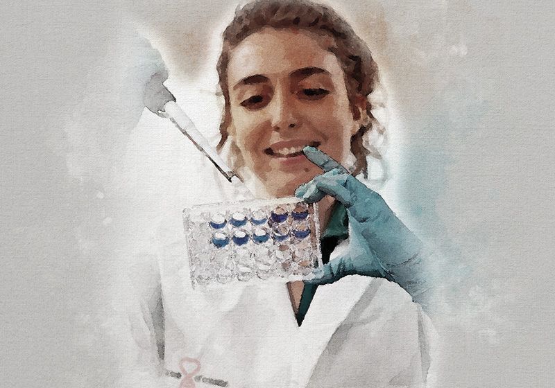
Cutting-edge spatial transcriptomics allows this postdoc to see where microbes live in the human body, how they behave, and what they mean for diseases.
Q | Write a brief introduction to yourself including the lab you work in and your research background.
I am Marta Reguera Gomez, a postdoctoral associate in the Frias-Lopez Lab at the University of Florida, where I study the spatial organization of the oral microbiome in human tissues. My background is in microbiology and infectious diseases, with expertise in parasitology, fungal diseases and, more recently, in metagenomics and host–microbe interactions.
Q | How did you first get interested in science and/or your field of research?
My interest in science began with a deep curiosity about how some organisms sustain themselves on others—the concept of parasitism. I was fascinated by the intricate strategies parasites have evolved to adapt to their hosts, to the point that their precision feels like high-tech design. For that reason, during my undergraduate studies in health biology, I joined a malaria research lab in Leiden, The Netherlands, and later continued with a master’s in tropical parasitic diseases at the University of Valencia, Spain. Since then, I have traversed almost the entire “pathogen spectrum,” working with helminths (worms), protozoa, fungi, and now bacteria. This journey has given me a broad perspective on the mechanisms that shape host–pathogen interactions and the delicate balance between survival and disease. It has also shown me that microbes are not just agents of infection, but essential players in human biology.
Q | Tell us about your favorite research project you’re working on.
One of my current research projects focuses on understanding how bacterial communities are spatially organized within human oral tissues. Traditional microbiome studies often rely on sequencing approaches that lose spatial context, leaving us with a list of microbes but little understanding of where they reside and how they interact with the host. In this project, I apply cutting-edge spatial transcriptomics technologies, such as 10x Genomics Visium, combined with advanced bioinformatics to map microbial presence directly within intact tissue sections. This approach allows me to visualize not only which bacteria are present, but also how they are organized among themselves and how they are positioned relative to host cells, immune infiltration, or diseased regions. I find this especially exciting because it bridges microbiology, pathology, and computational analysis, providing a holistic view of host–microbe interactions. Beyond the scientific novelty, I believe this work has strong translational potential, as spatially resolved microbiome data could help identify new diagnostic biomarkers or therapeutic targets for oral diseases and potentially other mucosal infections.
Q | What do you find most exciting about your research project?
Moving to the US from Spain has definitely been one of the most exciting (yet hard) experiences of my scientific journey so far. The cutting-edge technology available in US labs is unmatched, and it has allowed me to move my research to a higher level in both speed, efficiency, and quality. Beyond the technical advantages, the opportunity to collaborate with talented researchers from diverse backgrounds has expanded my scientific perspective and taught me to approach problems from multiple angles. At the same time, living and working abroad has pushed me to adapt, be resilient, and grow both professionally and personally. What I once saw as challenges (navigating a new system, culture, and language) have become some of the most rewarding parts of the journey. Ultimately, this experience has not only strengthened my scientific skills but also reaffirmed my passion for discovery and my commitment to contributing to science on a global stage.
Q | If you could be a laboratory instrument, which one would you be and why?
I would be a broad-spectrum fluorescent microscope. To me, it represents the perfect blend of science and art. With it, you don’t just generate numbers or abstract data, you witness living structures illuminated in vivid colors, moving, interacting, and creating patterns that feel almost like a painting in motion. Each image is both beautiful and deeply informative, revealing the architecture of cells and tissues while sparking a sense of wonder. A fluorescent microscope makes science feel tangible and alive, transforming complexity into something we can see with our own eyes, and that balance of creativity and insight is exactly how I like to approach research.
Are you a researcher who would like to be featured in the “Postdoc Portraits” series? Send in your application here.

