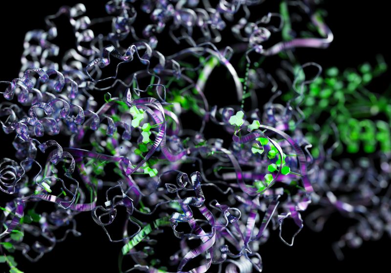
Proteins are most well-known for their intricate structures. The α-helices and β-sheets that form from interactions between sidechains create distinct shapes that, along with the specific amino acid sequences themselves, allow these molecules to perform unique functions in cells.
“Most proteins use all 20 amino acids—alanine, valine, methionine—they use all of them. So it’s like a card deck,” said Steven McKnight a molecular biologist at the University of Texas Southwestern Medical Center. Incorporating all or most of these amino acids creates complexity in the polypeptide chain that allows proteins to fold into their unique structures
However, some proteins contain regions, called low-complexity domains (LCDs), that are distinct from the highly organized helices and sheets of the molecules. “[These regions] don’t use the full deck of amino acids,” McKnight said. Instead, these LCDs only use a subset of the possible residues and remain more flexible. Without rigid structures, McKnight and other researchers struggled to study these disordered protein segments, making them the dark matter of protein biology.
Much like the enigmatic space stuff of the universe, LCDs are present in a surprising number of proteins, pointing to their vital functions in the cell.1 For example, they help form the barrier in nuclear pores that separate the nucleus from the cytoplasm. While researchers knew that nuclear pores consisted of an LCD that bound proteins for transport, they didn’t understand how this transport occured.2 Dirk Görlich, a biochemist at Max Planck Institute for Multidisciplinary Sciences, found himself wrapped up in this question.
Today (September 11), McKnight and Görlich won the 2025 Albert Lasker Basic Medical Research Award for their respective contributions to unraveling the structure and function of LCDs across the cell.
Elucidating the Function of LCD Repeats in Nuclear Pores
Eukaryotes perform DNA replication and transcription in the nucleus and protein synthesis in the cytosol, but materials for each function, like polymerases and RNA transcripts, need to be able to move between these two locations. Nuclear pores facilitate this transfer, selectively allowing these large materials to cross the nuclear membrane with the help of transporter proteins.
In the early 2000s, Görlich studied how nuclear pores formed this selective barrier. These nuclear pore proteins contained LCDs with a high abundance of paired phenylalanine (F) and glycine (G)-repeat domains that could bind the transporter proteins. Since this LCD had hydrophobic properties, many models at the time proposed that their function must revolve around repulsion.
“This was only half of the story,” Görlich said, adding that the other models did not explain how the pores selectively transported material or how the barrier formed.
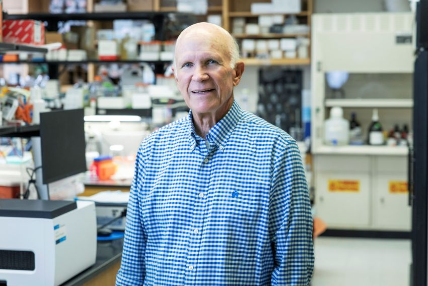
Dirk Görlich studies nuclear pore transport. He is a recipient of the 2025 Albert Lasker Basic Medical Research Award for his work studying the role of low-complexity domains in the nuclear pore.
Irene Böttcher-Gajewski, Max Planck Institute for Multidisciplinary Science
Instead, he suspected that transporter proteins were somehow suited to interact with the repeating FG domains to overcome its barrier function. “The key idea was that these low-complexity regions are not as non-interacting as people thought,” he said. He proposed a “selective-phase” model where FG-repeat domains in the nuclear pore interacted with each other to create a sieve-like structure that blocked macromolecules’ entry unless they could overcome this repulsion but that the transporter proteins could navigate. “There was a lot of resistance in our field [to the model],” he recalled.
To put the idea of these interactions to the test, Görlich and his team expressed nuclear pore FG-repeat domains in Escherichia coli and isolated them to determine if they would stay in an aqueous solution or engage with one another into a semi-solid structure. The FG repeats formed a millimeter scale hydrogel after being dissolved and exposed to physiological pH. The team showed that replacing the hydrophobic phenylalanine with a hydrophilic serine prevented gel formation. This demonstrated that these LCDs in fact interact and form a barrier.3
To further explore how this barrier selectively allowed for the passage of proteins, they first measured how fast proteins moved through a gel of FG repeats. “It was clear that we just had to measure three rates to describe the process,” Görlich said. They studied the rate of entry, diffusion, and exit through their hydrogel.
The team showed that this hydrogel efficiently and rapidly shuttled import proteins through the material while excluding large proteins not bound to a transport protein.4 “The numbers were surprisingly close to what we had measured with nuclear pores,” Görlich said.
In fact, Görlich and his team saw that proteins diffusing to the surface the gel represented the rate-limiting step, not entry into the gel. “It was like a black hole just attracting the stuff,” he recalled.
He added that, today, the rate of this passage and the ability to replicate it with the hydrogel stands out to him as the most impressive finding. “I would never have thought that we can have a material that absorbs in protein so rapidly that the diffusion through the water becomes rate-limiting,” he said.
Finally, Görlich and his team replicated these findings in nuclear pore complexes from frog eggs. They showed that removing the repeated FG domains or modifying hydrophobic residues to hydrophilic serines in different nucleoporins reduced the barrier function of the pore.5 Additionally, the selective permeability and efficient transporter roles of the pores were dependent on the ability of the FG repeats to interact with one another. “This was a clear contradiction to the other models [that] thought you don’t need an interaction between the FG repeats,” Görlich said.
Collectively, these findings supported Görlich’s selective phase model of the nuclear pore complexes. “It’s quite satisfying to see that very, very simple physics can describe the function of a super complex structure,” he said.
Recently, Görlich and his team demonstrated that they could replicate this barrier function using a peptide consisting of 12 amino acids, which includes an FG domain, that repeats 52 times.6 “We can reduce the complexity of the structure to a 12mer peptide, and this is a mind-blowing thing,” he said. This repeating polypeptide could help researchers explore how the barrier forms structurally and the relationship between the sequences and barrier function, which Görlich said was an outstanding challenge in the field.
While these findings offer valuable insights into the innerworkings of cell biology, they can also inform other areas of research. For example, Görlich recently collaborated with researchers studying HIV to investigate how the virus’s capsid manipulates itself to gain entry into the nucleus, a vital part of its life cycle. They showed that the amino acids on the surface of the capsid protein allows it to act like a transporter protein to interact with the repeating FG domains and pass through the nuclear pore.7
An Accidental Discovery of Low-Complexity Domains in Subcellular Structures
While Görlich was building evidence for the selective-phase model in nuclear pores, McKnight was studying gene regulation. He first encountered LCDs while studying transcription factors in the 1980s. He and his colleagues demonstrated that the ones with LCDs required these unstructured regions to function properly, but without a 3D structure, they struggled to pin down that role.8 “It was exciting. [My colleagues and I were] thinking, ‘Whoa, this is really, really cool,” McKnight recalled. “But we couldn’t crack it open.” By the 1990s, McKnight decided to move on from the strange, shapeless sequences.

Steven McKnight studies gene regulation. He is a recipient of the 2025 Albert Lasker Basic Medical Research Award for his work studying the role of low-complexity domains in subcellular structures.
UT Southwestern Medical Center
However, in 2010, an unintended finding brought him back to LCDs. He and others had observed that an isoxazole compound added to embryonic stem cells prompted them to become cardiomyocytes.9 To tease apart the molecular mechanism of this phenomenon, McKnight and his team added this miracle chemical to cell lysates of different mouse tissues and cell lines. Instead of finding a specific protein in their purification assay, they pulled out a score of them. “That’s a disaster,” McKnight said. “Normally when you find an observation like that, you quit. You say, ‘No, that’s garbage.’”
Out of curiosity, though, McKnight decided to investigate the hundreds of proteins. He immediately noticed that they all shared a common denominator: an LCD.
McKnight knew the pattern pointed to an important piece of the puzzle. Digging further, he and his team showed that their isoxazole chemical bound this LCD; that explained the massive number of protein hits.
They decided to focus their efforts on one of these proteins, called fused in sarcoma (FUS). While studying FUS, one researcher stored a purified sample of it in the fridge overnight. The next morning, the team made another unexpected observation. In place of the aqueous solution was a gel.
“I had never seen that happen before with any protein that I had worked with,” McKnight recalled. Much like exploring his “disaster” protein purification, McKnight launched into finding out what caused this change in the FUS protein.
The team showed that, just like the binding to the compound, this gel appeared because of the LCD.10 McKnight realized that he and his team could leverage this gel transition as an assay to study LCDs that he didn’t have 20 years earlier. “Once you have an assay, it’s like you have a hammer, and now you’re looking for nails,” McKnight said.
One nail that they tackled was FUS’s ability to interact with RNA granules, small structures of RNA and ribonucleoprotein present throughout the cell. Researchers didn’t know how they formed. Using their assay like a hammer, McKnight and his team showed that these LCDs in FUS allowed it to form RNA granules.6
Taking the finding a step further, McKnight put a section of the FUS gel under the electron microscope. “We saw these beautiful polymers or fibers,” McKnight recalled. Subsequently, he and his team performed X-ray diffraction and saw a pattern that measured exactly 4.7 Ångstroms, exactly the distance between two β strands, suggesting that the polymers joined through cross-β interactions.10 They supported this further by studying the structure with nuclear magnetic resonance, identifying the exact location of the polymer-forming residues.11
The finding caused a stir amongst researchers, McKnight recalled; cross-β structures were features of pathological proteins. “A lot of people in the field of biology thought, ‘Oh, Steve’s crazy. This can’t be right because he’s saying that this is β-β interaction, and the cross-β interaction that he’s talking about is pathological. It kills you. So, there’s no way that Steve’s ideas are going to be right,’” McKnight said.
However, as McKnight and his team demonstrated, an important aspect of the LCD polymers was their labile nature. These cross-β structures were destined to fall apart, distinguishing them from more pathological varieties of polymers that used similar, but more stable, binding. “If it had been held together permanently, like super glue, then the likelihood that this would have been biologically meaningful would have been diminished,” McKnight said.
Instead, these LCDs create temporary interactions that enable the formation of RNA granules, as well as other membrane-less, subcellular structures. Collaborating with other researchers, McKnight showed that LCDs aided in the formation of intermediate filaments and in regulating transcription by interacting with RNA polymerase II.12,13
In 2022, McKnight and his team demonstrated how distinct mutations identified in different neurological disorders changed how LCD regions self-associate.14 “We could understand how the change of one single amino acid residue caused the interaction to become much too tight,” he said.
Research Worth Recognition
McKnight and Görlich’s intrepid and thorough explorations into the mechanisms of LCDs demonstrated the critical and unique properties of these protein regions. Through their independent studies, researchers across cell biology domains now have more footholds to explore cell transport and subcellular functions.
Görlich said that receiving the Lasker Award is a great recognition of his and his team’s work, especially considering some of the headwinds they encountered early on from the field. “Maybe the message for others is you should not get irritated if your colleagues tell you, ‘I don’t believe it’,” Görlich said. “Just do your thing and ask which experiments need to be done to distinguish between models.”
Reflecting on his career journey and Lasker Award, McKnight said that it’s especially great recognition for the trainees in his team who made the discoveries.
McKnight emphasized that surprising observations drive science. “The fun of it is that you go tinker with things, and you say, ‘Oh, my God, it works that way. I would have never thought of that.’”
- Cascarina SM, Ross ED. Identification of low-complexity domains by compositional signatures reveals class-specific frequencies and functions across the domains of life. PLoS Comput Biol. 2024;20(5):e1011372.
- Radu A, et al. The peptide repeat domain of nucleoporin Nup98 functions as a docking site in transport across the nuclear pore complex. Cell. 1995;81(2):215-222.
- Frey S, et al. FG-rich repeats of nuclear pore proteins form a three-dimensional meshwork with hydrogel-like properties. Science. 2006;314(5800):815-817.
- Frey S, Görlich D. A saturated FG-repeat hydrogel can reproduce the permeability properties of nuclear pore complexes. Cell. 2007;130(3):512-523.
- Hülsmann BB, et al. The permeability of reconstituted nuclear pores provides direct evidence for the selective phase model. Cell. 2012;150(4):738-751.
- Ng SC, et al. Recapitulation of selective nuclear import and export with a perfectly repeated 12mer GLFG peptide. Nat Commun. 2021;12(1):4047.
- Fu L, et al. HIV-1 capsids enter the FG phase of nuclear pores like a transport receptor. Nature. 2024;626(800):843-851.
- Triezenberg SJ, et al. Functional dissection of VP16, the trans-activator of herpes simplex virus immediate early gene expression. Genes Dev. 1988;2(6):718-729.
- Sadek H, et al. Cardiogenic small molecules that enhance myocardial repair by stem cells. Proc Natal Acad Sci USA. 2008;105(16):6063-6068.
- Kato M, et al. Cell-free formation of RNA granules: Low complexity sequence domains form dynamic fibers within hydrogels. Cell. 2012;149(4):753-767.
- Murray DT, et al. Structure of FUS protein fibrils and its relevance to self-assembly and phase separation of low-complexity domains. Cell. 2017;171(3):615-627.e16.
- Sysoev VO, et al. Dynamic structural order of a low-complexity domain facilitates assembly of intermediate filaments. Proc Natl Acad Sci USA. 2020;117(38):23510-23518.
- Kwon I, et al. Phosphorylation-regulated binding of RNA polymerase II to fibrous polymers of low-complexity domains. Cell. 2013;155(5):1049-1060.
- Zhou X, et al. Mutations linked to neurological disease enhance self-association of low-complexity protein sequences. Science. 2022;377(6601):eabn5582.

