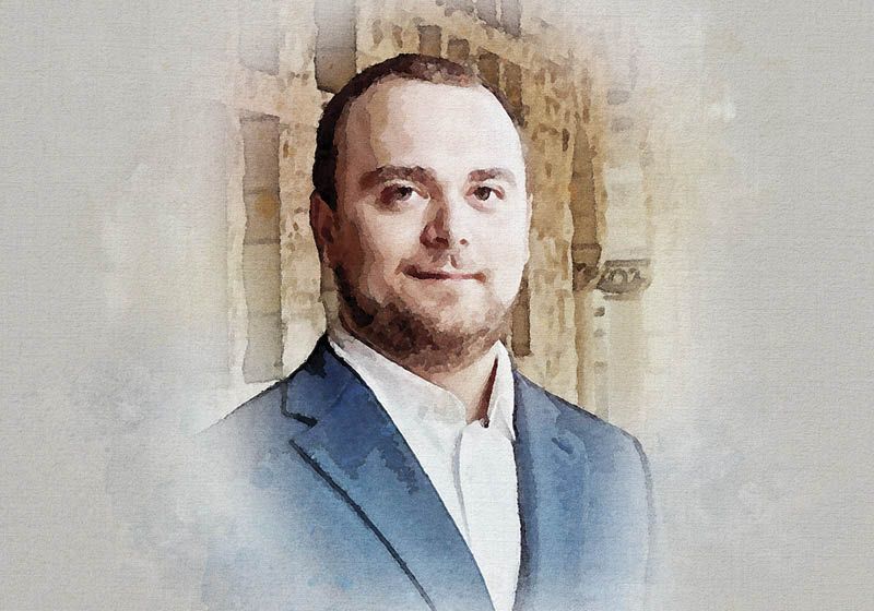
This physicist-turned-biomedical researcher is blending nanomaterials and imaging, in the quest for personalized medicine.
Q | Write a brief introduction to yourself including the lab you work in and your research background.
I am Giovanni Marco Saladino, a postdoctoral scholar in the Daldrup-Link lab at Stanford University. With a PhD in Biological and Biomedical Physics from the Royal Institute of Technology, my research background covers molecular imaging, nanomedicine, and nanomaterials, focusing on the development of multimodal contrast agents for theranostic (therapy + diagnostic) applications.
Q | How did you first get interested in science and/or your field of research?
From a young age, I was fascinated by the hidden structures and mechanisms that shape the natural world. This curiosity drew me first toward physics, where I learned how fundamental principles can explain complex phenomena. During my graduate studies, I became increasingly intrigued by how these principles could be applied to living systems, leading me to the field of biomedical physics. I was inspired by the possibility of using physical tools, such as imaging techniques and nanomaterials, to understand the physical phenomena at a deeper level and to directly impact human health. This motivation guided my PhD research on nanomaterials for imaging applications, followed by my postdoctoral work in molecular imaging and nanomedicine\. For me, the most rewarding aspect of science is the balance between creativity and rigor: the ability to imagine new solutions while grounding them in careful experimentation. The prospect of developing technologies that may one day improve diagnosis and therapy continues to drive my enthusiasm for research.
Q | Tell us about your favorite research project you’re working on.
One of my favorite projects focuses on developing multimodal nanomaterials for imaging and therapeutic applications. The idea is to engineer nanoparticles that can be simultaneously visualized with different imaging modalities, such as MRI and X-ray fluorescence imaging. This dual functionality allows us to monitor where the particles go in the body, assess how they interact with tissues, and ultimately track the effectiveness of treatments in real time with more accuracy. I particularly enjoy this project because it sits at the intersection of physics, chemistry, and medicine, requiring collaboration across disciplines. It is both conceptually challenging and deeply rewarding: every experiment provides not only data but also new questions about how to optimize design and translate findings into preclinical models. What excites me most is the long-term potential of this research to help clinicians personalize therapies and improve patient outcomes, which gives a strong sense of purpose to the technical challenges I work through every day.
Q | What do you find most exciting about your research project?
The most exciting part of my scientific journey has been witnessing the transition from abstract ideas to tangible results that can make a real difference in medicine. During my PhD, I worked on designing nanoparticles for imaging, and I still remember the first time I saw clear experimental data confirming that the particles behaved exactly as I had expected. That moment captured the essence of why I enjoy research: the mix of creativity, persistence, and discovery. Equally exciting has been the opportunity to collaborate with scientists from diverse fields, each bringing unique expertise and perspectives. These collaborations have not only strengthened the science but also expanded my own way of thinking, teaching me to view challenges from multiple angles. Now, as a postdoctoral scholar, I find great motivation in advancing molecular imaging technologies toward clinical relevance. Knowing that the work I do could one day improve how diseases are detected and treated remains the most rewarding and inspiring aspect of my career so far.
Q | If you could be a laboratory instrument, which one would you be and why?
I would be an MRI scanner. I admire how MRI combines physics, engineering, and medicine to create images that reveal what is otherwise invisible. Like an MRI, I enjoy looking beneath the surface to uncover hidden layers of meaning and possibility. MRI also requires patience and precision, characteristics I strive to bring to my own work. At the same time, it is versatile. With the right adjustments, MRI can provide structural, functional, and molecular information. I value that same flexibility in my career, where I enjoy moving across disciplines and adapting to new challenges. Most importantly, MRI is noninvasive, reflecting my own belief that science should seek to illuminate and improve without causing harm. For me, the MRI symbolizes curiosity, rigor, and impact, making it the perfect instrument to embody.
Are you a researcher who would like to be featured in the “Postdoc Portraits” series? Send in your application here.

