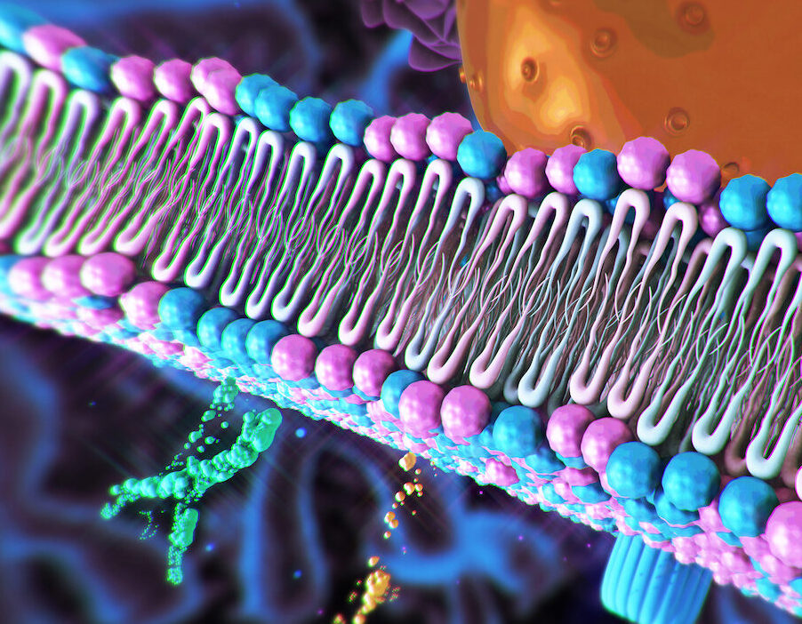
Cellular membrane proteins play important roles in cellular transport, signaling, and cell-to-cell communication. Malfunction in membrane proteins can lead to serious diseases, such as cancer. However, current biophysical models poorly describe the behaviors and energetics of proteins within lipid bilayers, limiting the understanding of disease-related misfolding and mechanisms for therapeutic intervention.
In a new study published in PNAS titled “Design principles of the common Gly-X6-Gly membrane protein building block,“ researchers from Scripps Research have unveiled a protein design approach for decoding the sequence–structure relationships underlying transmembrane α-helix packing. The findings offer computational strategies to interpret and manipulate membrane protein folding and function for therapeutic targeting and more biotechnological applications.
“Billions and billions of dollars a year are going into making molecules that target membrane proteins to alter their behavior and combat disease, but in order to modulate these proteins, it helps to first understand how they work,” said Marco Mravic, PhD, an assistant professor in the Department of Integrative Structural and Computational Biology at Scripps Research and corresponding author of the study. “Our study uncovered some new rules of sequence and atomic arrangements inside membrane proteins that are essential for them to function.”
Membrane proteins consist of multiple helices that are folded and packed tightly together. To maintain their complex architecture and function, different parts of the protein must bind more tightly to each other than to the lipid membrane in which they’re embedded.
The researchers hypothesized that common structural motifs represent potential “sticky” spots to assist membrane protein helices in binding to each other and organizing within their membrane folds.
“It’s usually really hard to study how membrane proteins behave within our bodies, because as soon as we break them out of the cell, they want to fall apart,” said Mravic. “Our approach is unique in that we design new synthetic proteins from scratch with computer programs to approximate the behaviors and atomic structures of membrane proteins from nature. We can use these designer proteins as models to ask questions and clarify rules underlying many of the complex processes happening within cell membranes that we could not see or study otherwise.”
Kiana Golden, first author of the study, developed a computational pipeline to identify protein sequences that folded onto the desired motif and designed optimized synthetic membrane proteins with enhanced stability. Sequences were experimentally validated to fold into the motif as predicted, supporting the hypothesis that these motifs create “sticky spots” between adjacent helices that hold the membrane proteins together in lipid. Additionally, newly designed sequences showed strong stability and even remained intact under boiling conditions.
“We found that the motif’s stability was driven by an unusual type of hydrogen bond that’s typically very weak, but when the motif is repeated, these weak hydrogen bonds all add up to make a very stable interaction,” said Golden, who worked on the project as a UCSD undergraduate and is now a graduate student at Princeton University. “This type of hydrogen bond is rare in the natural world, so it was really surprising that this is largely what’s driving this motif to occur, and that biology has evolved to use it within specific motifs and structures across nature.”
Understanding how this motif supports membrane protein structure offers a path to identify genetic mutations that could contribute to disease. In future work, the research team will design molecules to directly target membrane proteins within the cell.
“Our approach vastly accelerates what we can discover about the inner workings of membrane proteins and how to make better therapies,” said Mravic.

