Our cells are continually being exposed to environmental assaults. For instance, skin is exposed to pollutants and UV radiation. Our lungs are subject to environmental stimuli such as smoke particulates and viral agents. Collectively, these environmental assaults can be summed as “exposomes”—the non-genetic factors that affect the health and biology of our tissues. In particular, our immune system is extremely sensitive to exposomes, which can drive the body into inflammatory states.
Models that most resemble human physiology are needed to fully understand and characterize the health impact of exposomes. The traditional models are 2D cell culture, 3D spheroids, and animals, but they have their respective limitations. 2D cell culture neglects the influence of 3D structure and extracellular matrix, while 3D spheroids lack sufficient cell diversity. Animals do not have the identical cell types as humans, and even for humanized mice models, it is often a mix of human and animal cell types. As a result, preclinical data obtained using these models often do not lead to meaningful outcomes during clinical trials.
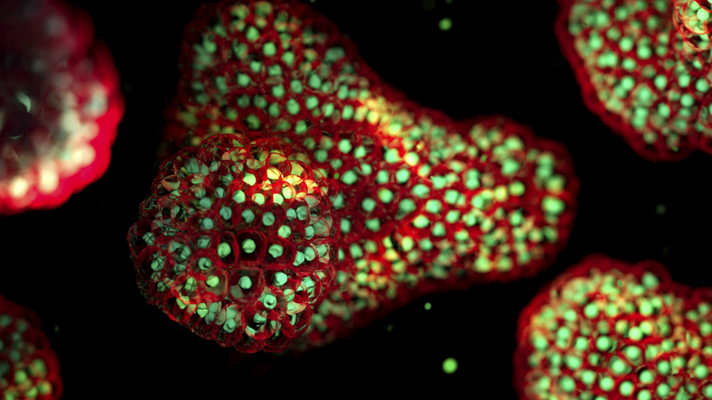
Organoids are 3D self-organized tissue structures that mimic critical structural and functional aspects of organs.1 While not perfect, they possess key features such as high cell diversity, structural similarities, and human origins which are making them increasingly popular models for understanding the impact of exposomes on human biology. The recent announcement by the U.S. Food and Drug Administration (FDA) to phase out animal testing requirements for therapeutic evaluation is also motivating researchers to develop more physiologically authentic organoid systems.
What follows are descriptions of some of the recent progress in using organoids for exposome research, especially in the use of artificial intelligence (AI) for data analysis.
Although organoids of various bodily systems were developed more than two decades ago, the first immune organoid was reported only in 2021, and again more recently in 2024, by two separate groups. This delay is because most organoids can be derived by differentiating induced pluripotent stem cells (iPSCs) into the desired cell lineage, but differentiation of iPSCs to lymphoid cell lineages such as B cells and T cells is not highly effective. This fact also meant that the percentage of cell population would be heterogenous and poorly controlled, and this would introduce inconsistencies during assay readouts.
Furthermore, through iPSC technology, the immune memory of the donor would be erased and the advantage of donor-specific drug screening for personalized medicine would be lost. This means that creating immune organoids requires a drastically different approach than to conventional organoids.
Donors’ immune memory retained
In 2021, Lisa Wagar, PhD, and colleagues at Stanford University developed the first immune organoids by digesting tonsil tissues and seeding them into transwells.2 They found that their organoids were able to retain 16 different immune cell types including B and T cells, which were difficult to obtain from iPSCs. Immunization of their organoids also elicited memory recall response with flu-specific antibodies, demonstrating that donors’ immune memory was retained.
Using B-cell receptor sequencing, the team also discovered that their organoids were able to support somatic hypermutation, a process in which the body rapidly mutates antibody genes in the variable region of immunoglobulin genes of activated B cells to produce a wide array of antibodies. This trait was achieved by administering virus exposomes to the organoid culture.
Nevertheless, in this transwell organoid model, the tissue microenvironment was missing. Thus, the immune organoid looked like a loose aggregate of cells, which made downstream analyses such as tissue slicing technically challenging. Furthermore, to use organoids to study the impact of exposomes in a high throughput manner, it is necessary for the organoids to be made reproducibly. In late 2024, Zhong and colleagues at the Georgia Institute of Technology reported the second immune organoid model by mixing hydrogel biomaterials with tonsil-derived cells.3 They first measured the mechanical properties of tonsil tissues before optimizing the synthetic hydrogel using four-arm poly(ethylene glycol) macromer end-functionalized with maleimide groups (PEG-4MAL), conjugated to cysteine-flanked, VCAM-1-mimicking REDV peptides.
In their model, germinal centers were more clearly visible compared to the first paper, but the hydrogel degraded rapidly, approximately 60% by day four. Hence, a human dermal fibroblast cell line was also added to the organoid model to maintain the extracellular matrix. However, this cell line was non-donor matched, which could introduce immune mismatch and attack, although this was not explained or studied. In patients with lymphoma, the team found that their organoids were only able to mount a weak immune response against influenza exposome stimulation.
“The human organoid system is particularly important in infectious disease research. Beyond serving as a physiologically relevant model to study viral pathogenesis, it has recently been applied to recapitulate key germinal center features in vitro, enabling the evaluation of humoral immune responses to vaccines without reliance on animal models—which are often poorly predictive of human vaccine responses,” says Chee Wah Tan, PhD, research assistant professor at the National University of Singapore.
AI-enabled heart organoid analysis
Cardiovascular disease is the leading cause of death worldwide, and it is associated with heart inflammation, which can cause severe cardiac dysfunction due to damage from cytokines, including interferon-γ and tumor necrosis factor-α. Inflammation also affects the cells in heart organoids, depending on the cytokine concentration and how long they are being exposed to inflammatory assaults. It is crucial to develop a technique that can measure inflammation rapidly using heart organoids.

A team led by Haotian Bai, PhD, and Shu Wang, PhD, at the Chinese Academy of Sciences, built a database containing videos on how cytokines at different concentrations and time points affected heart organoids’ morphologies and functions.4 They then used an AI algorithm based on gradient boosting—called XGBoost—to extract key features and quantify the correlation between experimental parameters and assay readouts. The team found that treatment with 10 ng mL−1 of interleukin-1β caused the most severe heart inflammation in their AI model.
Further validation was performed with single-cell RNA sequencing, which found that cardiac inflammation disproportionately led to the death of endothelial cells. Finally, to treat heart inflammation, they extracted nanothylakoid units from plants and gave them to the heart organoids (nanothylakoid supplied adenosine triphosphate (ATP) and nicotinamide adenine dinucleotide phosphate (NADPH) to reduce oxidative stress and inflammation-related injuries).
“Developing cardiac inflammatory organoid models incorporating multiple cell lineages is vital for novel therapies. Our motivation for developing this technology stems from three critical needs in cardiac inflammatory organoid research: (1) Significant variability in the types, induction timing, and concentrations of inflammatory mediators in constructing cardiac inflammatory organoids; (2) Overreliance on complex, low-throughput evaluation methods, and (3) Narrow assessment dimensions that fail to capture inflammation’s multifaceted impact,” says Bai, professor at the Chinese Academy of Sciences.
“We hypothesized that revealing the morphological and functional changes of heart organoids caused by inflammation can improve our understanding of the effects of inflammation on the heart. In this paper, we also address analytical challenges with AI as current manual analysis struggles with organoids’ small size, high heterogeneity, and multi-dimensional pathological changes. We aim to enable rapid, quantitative assessment of heart organoids to accelerate therapeutic development.
“Our platform uniquely integrates brightfield video–based radiomics with interpretable machine learning to enable rapid, quantitative scoring of inflammation in cardiac organoids. By extracting morphological, textural, and functional features from time-lapse imaging, the system generates objective, reproducible metrics within 30 minutes. Compared with conventional organoid evaluation models—often dependent on labor-intensive biochemical assays—our approach is faster, non-destructive, and highly scalable.”
Wang, also a professor at the Chinese Academy of Sciences, adds that “AI is central to transforming raw brightfield microscopy videos into actionable biomarkers. Automated image segmentation and high-dimensional radiomic feature extraction detect subtle morphological, textural, and functional changes in organoids. Interpretable machine learning models link these features to inflammation severity and therapeutic response. This allows real-time, non-destructive monitoring of treatment effects, significantly shortening evaluation timelines, increasing throughput, and improving the predictive value of organoid-based drug screening.”
The two colleagues agree that their current work still suffer from some limitations: (1) further optimization is needed for cardiac organoid construction; (2) reliance on commercial stem cells limits the ability to model patient-specific conditions, hindering personalized drug development; (3) the algorithm requires iterative upgrades to meet commercial software standards; and (4) application scenarios remain narrow, with limited use across different exposome testing contexts.
To address these issues, they plan to collaborate with hospitals to generate patient-specific cardiac organoids using reprogrammed stem cells, optimizing cytokine combinations to improve culture quality. In parallel, they will refine their algorithm using large-scale compound testing data from clinical patient samples, enabling the development of commercial-grade software for more accurate, convenient, and individualized in vitro exposome testing.
“We are enhancing our intelligent algorithm to improve accuracy, drawing on data from hospital-derived cardiac organoids exposed to diverse environmental factors. In parallel, we are developing a user-friendly imaging software solution: users will simply photograph the heart organoids according to guidelines provided, and the software will automatically analyze the images and rapidly deliver exposome testing results. We are already working on these developments and look forward to sharing the latest progress with readers soon,” says Bai.
AI to improve drug screening using cancer organoids
Pancreatic ductal adenocarcinoma (PDAC) accounts for more than 90% of all pancreatic malignancies and is one of the deadliest cancer types. Not only is its incidence increasing but it also a highly heterogenous cancer with several key cancer gene drivers, making it challenging to identify a single therapeutic molecule that works for all PDAC patients.
Unfortunately, PDAC is also surrounded by a thick dermal barrier that prevents immune cells from entering the tumors, making this cancer type an immune-excluded or -desert form. Technologies that can enable unbiased and systematic analysis of T cell-mediated tumor recognition on an individual patient basis would greatly contribute to predicting whether an individual patient will be sensitive to immunotherapy.
Nathalia Ferreira, a PhD student, and colleagues developed patient-derived PDAC organoids and developed OrganoIDNet, an AI algorithm that uses live cell imaging data to assess organoid growth and functions, and the efficacy of immunotherapy.5 The authors argue that this strategy allows accurate monitoring of the longitudinal response of PDAC organoids to chemotherapy in organoid cultures and immunotherapy in a novel organoid co-culture set-up with peripheral blood mononuclear cells (PBMCs).
In their paper, they added PD-L1 inhibitor (Atezolizumab) before using OrganoIDNet to identify how the drug affected organoid size, eccentricity, and T-cell infiltration and cytotoxicity. The team says that their approach will support the pre-clinical testing of personalized immunotherapy-based treatment strategies for PDAC patients.
Organoids are promising in vitro models that can be used to mimic human physiology and provide accurate information on how human cells in their native-like tissue microenvironment are affected by exposomes including pollutants, toxic metal ions, drugs, viruses, and even microplastics. The development of immune organoids is expected to accelerate the synthesis of assembloid models in which environmental exposomes trigger immune-organ crosstalk. Furthermore, with rapid progress in AI methods, downstream analyses of organoid health and functions should become more precise and common, paving the way for organoids to make a larger translational impact on biomedical innovations.
Andy Tay, PhD, is a Presidential Young professor in the department of biomedical engineering at the at the National University of Singapore (NUS) and principal investigator at the Institute of Health Innovation & Technology and the NUS Tissue Engineering Program. He is also a freelance science journalist.
References
- Zhao Z, Chen X, Dowbaj AM, et al. Organoids. Nature Reviews Methods Primers. 2022;2(1). doi:10.1038/s43586-022-00174-y
- Wagar LE, Salahudeen A, Constantz CM, et al. Modeling human adaptive immune responses with tonsil organoids. Nat Med. 2021;27(1):125-135. doi:10.1038/s41591-020-01145-0
- Zhong Z, Quiñones-Pérez M, Dai Z, et al. Human immune organoids to decode B cell response in healthy donors and patients with lymphoma. Nat Mater. Published online 2024. doi:10.1038/s41563-024-02037-1
- Lin H, Cheng J, Zhu C, et al. Artificial Intelligence-Enabled Quantitative Assessment and Intervention for Heart Inflammation Model Organoids. Angewandte Chemie – International Edition. 2025;64(25). doi:10.1002/anie.202503252
- Ferreira N, Kulkarni A, Agorku D, et al. OrganoIDNet: a deep learning tool for identification of therapeutic effects in PDAC organoid-PBMC co-cultures from time-resolved imaging data. Cellular Oncology. 2025;48(1):101-122. doi:10.1007/s13402-024-00958-2

