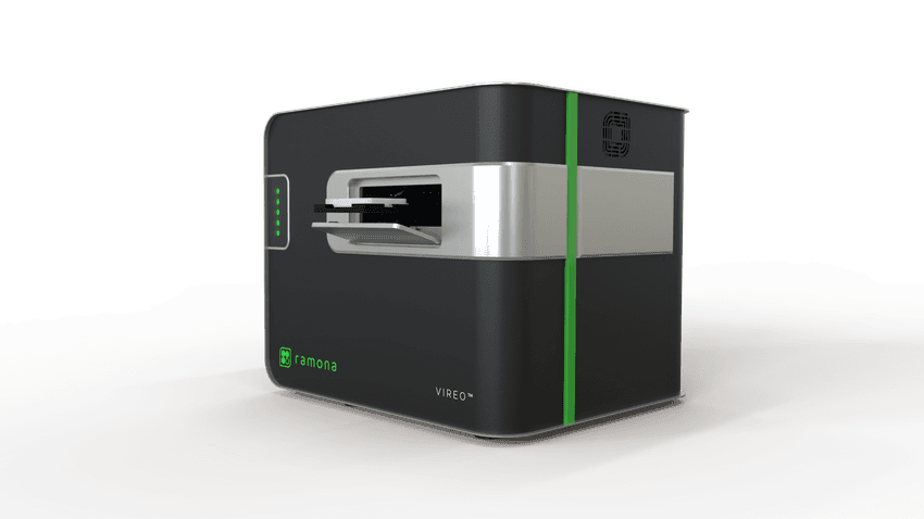
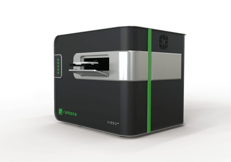
The Vireo platform from Ramona brings unprecedented speed to live-cell imaging with the help of multi-camera microscopy.
Researchers seeking to explore live-cell dynamics can now turn to rapid, precise, and gentle technologies that address conventional microscopy challenges such as limited temporal resolution and photodamage risk. Ramona Optics’ live-cell imaging platform, Vireo, replaces slow single-view snapshots with parallelized video microscopy, quickly capturing multi-sample functional insights across whole 96-well plates.
“We’re really trying to take the process of image acquisition, for many samples at once, from hours on other systems down to minutes and even seconds,” explained Natalie Alvarez, product lead at Ramona. “Vireo essentially combines 24 microscopes into one compact benchtop system, and that enables us to deliver unprecedented scale with regards to high-throughput screening as well as live functional drug studies or interaction studies among cells and organoids.”
Built on Ramona’s multi-camera array microscopy (MCAM) architecture, Vireo packs multiple 4K video microscopes and content-aware optics into a compact plate reader that captures brightfield and multi-channel fluorescence at unprecedented speed and scale. Its unique parallel imaging and automated z-stacking with low-dose illumination capabilities help preserve cellular resolution while multiplying temporal sampling and reducing photodamage, enabling whole-plate scans in roughly one to two minutes.

Vireo combines the power of 24 miniaturized microscopes and content-aware optics into a compact plate reader that captures live-cell data at unprecedented speed and scale.
Ramona Optics, Inc.
One area where Vireo is already making its mark is by helping scientists characterize dynamic biological processes in 3D cultures, enabling researchers to see previously unviewable processes at high-throughput speeds. “We are looking into neural psychiatric disorders, and these have been inherently hard to model, mostly because we don’t have access to living human brain tissue. Brain organoid technology combined with the ability to do live-cell imaging is breaking that barrier,” said Katharina Meyer, a senior scientist in the Synthetic Biology Platform at Harvard University’s Wyss Institute.
Meyer first accessed Vireo through the Wyss Institute’s scientific instrumentation program, which connects industry partners with scientists to drive innovation and new discoveries. She is currently implementing Vireo in a high-throughput workflow for optimal brain organoid culture quality control testing in a 96-well environment. “This has been ultra-fast. Within a few seconds, we have imaged an entire plate and can produce the data that we need to phenotype the organoid, such as size, density, and shape,” Meyer explained. “That we have the ability to look at the signaling context in a live brain-like environment, this is what is really exciting about this, and also that it’s done so quickly.”

