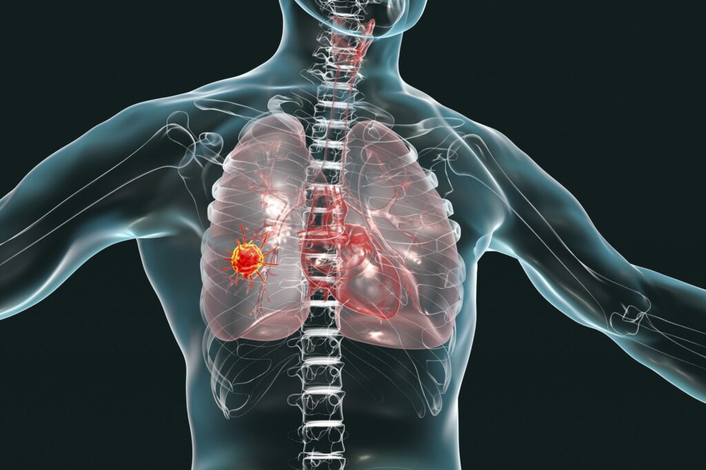
Lung cancer remains the world’s deadliest malignancy, largely because most tumors are discovered only after they have spread. For years, scientists have sought molecular clues that precede tumor formation, hoping to uncover a window for early intervention. In a new study, published in Cancer Cell, MD Anderson researchers combined spatial transcriptomics with molecular pathology to visualize how lung cells transition from healthy to precancerous to malignant.
By integrating data from 56 human precursor and advanced tumor samples across 25 patients and validating their results in an additional 36 lesions from 19 patients, the team built a cellular atlas containing more than five million cells. The resulting maps capture the architecture of early lesions with unprecedented clarity, showing precisely where cancer-initiating cells appear and what signals surround them.
“We find that the earliest cells that give rise to lung cancer are in regions with very high inflammation and are surrounded by proinflammatory cells,” said Humam Kadara, PhD, professor of Translational Molecular Pathology at MD Anderson. “Targeting inflammation by neutralizing a driver called IL-1B reduces these precursor cells of lung cancer. Our work paves the way for targeting inflammation to intercept the earliest stages of lung cancer and impact patient lives.”
Mapping inflammation at single-cell resolution
Spatial transcriptomic analysis allowed the researchers to determine not only which genes were active but where they were turned on. In precursor lesions, the team identified clusters of alveolar epithelial cells that had begun to acquire early tumor-like features. These cells were consistently embedded within zones of intense inflammatory activity rich in macrophages, neutrophils, and T cells expressing cytokines such as IL-1B and TNF-α.
The proinflammatory regions were strikingly more abundant in early lesions than in advanced tumors, suggesting that inflammation is not a by-product of cancer but a precursor condition that promotes its emergence. Laboratory models reproduced the same pattern: suppressing IL-1B signaling reduced the number of pre-malignant cells, directly linking inflammatory cues to tumor initiation.
Defining the earliest molecular transitions
The data also revealed how gene-expression programs evolve as lung lesions progress. In early stages, epithelial cells upregulated pathways related to stress responses, interferon signaling, and cell adhesion—changes consistent with an environment under chronic immune attack. As lesions advanced, the cells gradually activated canonical oncogenic networks involving EGFR, KRAS, and p53, suggesting that inflammation may create the mutagenic and selective pressures that give rise to these driver mutations.
Importantly, even before overt malignancy, immune-checkpoint molecules such as PD-L1 and IDO1 were detected within inflammatory niches, implying that the earliest tumor precursors already engage immune-evasion mechanisms. These insights help explain why many lung cancers are resistant to immunotherapy once established, and why combining anti-inflammatory agents with checkpoint blockade could improve outcomes.
From spatial biology to clinical interception
Co-senior author Linghua Wang, MD, PhD, professor of Genomic Medicine and associate member of the James P. Allison Institute, emphasized that spatial transcriptomic mapping provides more than static images; it exposes the dynamic interactions between cells. By reconstructing how inflammatory signals sculpt the microenvironment over time, the team has identified molecular targets that may prevent premalignant lesions from ever becoming invasive cancer.
Spatial mapping also uncovered biomarkers that could guide risk stratification in lung-screening programs. Certain proinflammatory gene signatures, particularly high IL-1B and NLRP3 inflammasome activity, were enriched in lesions that later progressed. Detecting these markers in sputum or minimally invasive biopsies might help identify patients who would benefit from anti-inflammatory or immune-modulating interventions.
The identification of IL-1B as a critical node in early tumor initiation resonates with prior population studies. In the CANTOS trial, patients receiving the IL-1B–blocking antibody canakinumab for cardiovascular disease had a markedly lower incidence of lung cancer—a finding that now gains mechanistic support. Kadara’s team directly demonstrated that neutralizing IL-1B in experimental models reduced the pool of precursor cells, reinforcing the concept that chronic, unresolved inflammation can act as an oncogenic engine.
Clinical implications and next steps
The findings converge on a new paradigm: lung cancer may be less a sudden genetic accident and more the cumulative outcome of persistent inflammatory signaling in vulnerable epithelial cells. If inflammation is indeed the spark, extinguishing it early could prevent ignition.
The MD Anderson group envisions future trials testing IL-1B or NF-κB pathway inhibitors as chemopreventive agents in high-risk individuals, such as heavy smokers or patients with chronic obstructive pulmonary disease, where lung inflammation is common.
The study also provides a roadmap for integrating spatial biology into precision prevention, combining imaging, molecular profiling, and computational modeling to track how tissues evolve toward malignancy.

