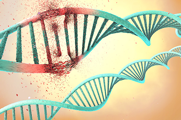
A new fluorescent sensor is giving scientists an unprecedented view of how cells respond to DNA damage, capturing the repair process as it unfolds in real time. The tool, developed at Utrecht University and published in Nature Communications, could accelerate research in cancer biology, drug safety testing, and aging. The paper is titled “Engineered chromatin readers track damaged chromatin dynamics in live cells and animals.”
Our DNA is constantly under assault from sunlight, chemicals, radiation, and even normal metabolic processes. While cells usually repair this damage quickly, failures in repair can contribute to cancer, neurodegeneration, and aging, and we know that a lowered capacity for DNA repair is linked to many human diseases. Until now, however, researchers have struggled to watch repairs directly. Most methods relied on antibodies that bind to damaged DNA, but these approaches required fixing and killing cells at different time points and can interfere with the cell’s ability to repair itself, offering only static snapshots.
The Utrecht team engineered a live-cell sensor that changes this picture entirely. Built from the tandem BRCT domain of the protein MCPH1, the probe binds briefly to the histone mark γH2AX, which appears at sites of DNA double-strand breaks. By attaching a fluorescent tag, the researchers created a tool that lights up DNA damage without interfering with the cell’s own repair machinery.
“Our sensor is different,” explained lead researcher Tuncay Baubec, PhD. “It’s built from parts taken from a natural protein that the cell already uses. It goes on and off the damage site by itself, so what we see is the genuine behavior of the cell.”
That dynamic binding is key. Unlike antibodies or nanobodies that can block repair proteins, the MCPH1-based sensor associates and dissociates rapidly, allowing scientists to track the full kinetics of repair. In live-cell imaging experiments, the probe revealed how damage foci form within minutes of exposure to genotoxic agents such as etoposide or ultraviolet light, and how they resolve over hours as repair progresses.
Richard Cardoso Da Silva, PhD, who engineered and tested the tool, recalls the moment he realized its potential. “I was testing some drugs and saw the sensor lighting up exactly where commercial antibodies did,” he said. “That was the moment I thought: this is going to work.”
The researchers can now see when damage appears, how quickly repair proteins arrive, and when the cell finally resolves the problem. “You get more data, higher resolution and, importantly, a more realistic picture of what actually happens inside a living cell,” said Cardoso Da Silva.
The team also demonstrated the sensor’s versatility beyond cultured cells. In the nematode C. elegans, the probe revealed programmed DNA breaks that occur naturally during gametogenesis. This suggests broad applicability in living organisms, not just cell lines.
Although the sensor itself is not a therapy, its potential impact on translational research could be useful moving forward. Many cancer treatments rely on inducing DNA damage in tumor cells, and aging studies increasingly focus on repair capacity. By making the probe openly available, the Utrecht team hopes to accelerate discoveries across fields.
With this innovation, researchers can finally watch DNA fix itself—live, in real time—opening a new window into one of biology’s most fundamental processes.

