Researchers at Duke-NUS Medical School have developed a detailed single-cell atlas of the developing human brain, offering a new reference point for evaluating lab-grown neurons used in Parkinson’s disease (PD) research and other brain disorders. The study, titled “BrainSTEM: A single-cell multi-resolution fetal brain atlas reveals transcriptomic fidelity of human midbrain cultures,” and published in Science Advances, introduces a two-tier mapping framework called BrainSTEM (Brain Single-cell Two tiEr Mapping), which helps assess how closely in vitro models resemble actual midbrain development.
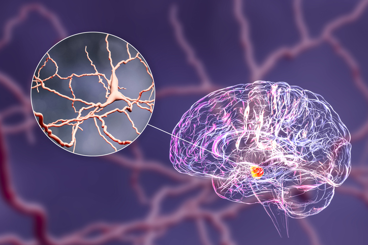
The team analyzed nearly 680,000 cells from fetal brain tissue spanning postconception weeks three to 14 from two datasets. The data generated was then used to build two reference atlases: one covering the entire brain and another focused specifically on the midbrain. The midbrain is of particular interest because it contains dopaminergic neurons—cells that produce dopamine and are affected in Parkinson’s disease.
BrainSTEM works by first projecting query datasets onto the whole-brain atlas to determine their regional identity. Cells identified as midbrain-like are then mapped onto the midbrain subatlas for more detailed analysis. “This second higher-resolution projection identifies mDA neurons as well as rare subpopulations that may otherwise be masked by nonmidbrain region cell types,” the authors wrote.
“Our data-driven blueprint helps scientists produce high-yield midbrain dopaminergic neurons that faithfully reflect human biology,” said co-first author Hilary Toh, an MD-PhD candidate at the Neuroscience & Behavioral Disorders program at Duke-NUS Medical School. Grafts of this quality are pivotal to increasing cell therapy efficacy and minimizing side effects, paving the way to offer alternative therapies to people living with Parkinson’s disease.”
The researchers evaluated 12 publicly available datasets along with their own in-house data, grouping them into two categories: (i) in vitro differentiation time series and (ii) PD models. They found that midbrain purity varied widely, with some cultures containing more than 50% cells from other brain regions. The study also showed that both 2D and 3D culture methods have limitations, and that “while the transition from radial glia to neuronal cells was observed in most developmental and differentiation time series datasets, the distribution of different neuronal subtypes varied across studies,” added the authors.
John Ouyang, PhD, a senior author, noted that BrainSTEM provides a way to benchmark these protocols more rigorously. “By mapping the brain at single-cell resolution, BrainSTEM gives us the precision to distinguish even subtle off-target cell populations. This rich cellular detail provides a critical foundation for AI-driven models that will transform how we group patients and design targeted therapies for neurodegenerative diseases.”
The atlas also identified previously uncharacterized subpopulations of midbrain cells and mapped their developmental trajectories. This could help researchers refine protocols to produce more targeted cell types for use in research or therapy.
While the study focuses on Parkinson’s disease, the BrainSTEM framework may be applicable to other neurological conditions. The researchers plan to make the atlas and mapping tools openly available, allowing other labs to use them for protocol evaluation and further study.
“BrainSTEM marks a significant step forward in brain modeling,” added assistant professor Alfred Sun, PhD. As researchers continue to explore its applications, the atlas may help accelerate progress in Parkinson’s research and contribute to more effective, targeted treatments for those affected by neurodegenerative conditions.

