Gidziela, A. et al. A meta-analysis of genetic effects associated with neurodevelopmental disorders and co-occurring conditions. Nat. Hum. Behav. 7, 642–656 (2023).
Buescher, A. V. S., Cidav, Z., Knapp, M. & Mandell, D. S. Costs of autism spectrum disorders in the United Kingdom and the United States. JAMA Pediatr. 168, 721–728 (2014).
Leigh, J. P. & Du, J. Brief report: Forecasting the economic burden of autism in 2015 and 2025 in the United States. J. Autism Dev. Disord. 45, 4135–4139 (2015).
Molnár, Z. et al. New insights into the development of the human cerebral cortex. J. Anat. 235, 432–451 (2019).
Wallace, J. L. & Pollen, A. A. Human neuronal maturation comes of age: cellular mechanisms and species differences. Nat. Rev. Neurosci. 25, 7–29 (2024).
Satterstrom, F. K. et al. Large-scale exome sequencing study implicates both developmental and functional changes in the neurobiology of autism. Cell 180, 568–584.e23 (2020).
Willsey, H. R., Willsey, A. J., Wang, B. & State, M. W. Genomics, convergent neuroscience and progress in understanding autism spectrum disorder. Nat. Rev. Neurosci. 23, 323–341 (2022).
Petilla Interneuron Nomenclature Group. Petilla terminology: nomenclature of features of GABAergic interneurons of the cerebral cortex. Nat. Rev. Neurosci. 9, 557–568 (2008).
Zeng, H. & Sanes, J. R. Neuronal cell-type classification: challenges, opportunities and the path forward. Nat. Rev. Neurosci. 18, 530–546 (2017).
Colonna, M. et al. Implementation and validation of single-cell genomics experiments in neuroscience. Nat. Neurosci. 27, 2310–2325 (2024).
Zeng, H. What is a cell type and how to define it? Cell 185, 2739–2755 (2022).
Yao, Z. et al. A high-resolution transcriptomic and spatial atlas of cell types in the whole mouse brain. Nature 624, 317–332 (2023).
Zhang, M. et al. Molecularly defined and spatially resolved cell atlas of the whole mouse brain. Nature 624, 343–354 (2023).
Langlieb, J. et al. The molecular cytoarchitecture of the adult mouse brain. Nature 624, 333–342 (2023).
Liu, H. et al. Single-cell DNA methylome and 3D multi-omic atlas of the adult mouse brain. Nature 624, 366–377 (2023).
Zu, S. et al. Single-cell analysis of chromatin accessibility in the adult mouse brain. Nature 624, 378–389 (2023).
Siletti, K. et al. Transcriptomic diversity of cell types across the adult human brain. Science 382, eadd7046 (2023).
Bhaduri, A. et al. Outer radial glia-like cancer stem cells contribute to heterogeneity of glioblastoma. Cell Stem Cell 26, 48–63.e6 (2020).
Smith, K. S. et al. Unified rhombic lip origins of group 3 and group 4 medulloblastoma. Nature 609, 1012–1020 (2022).
Li, M. et al. Integrative functional genomic analysis of human brain development and neuropsychiatric risks. Science 362, eaat7615 (2018).
Zhu, Y. et al. Spatiotemporal transcriptomic divergence across human and macaque brain development. Science 362, eaat8077 (2018).
Emani, P. S. et al. Single-cell genomics and regulatory networks for 388 human brains. Science 384, eadi5199 (2024).
Bhaduri, A. et al. An atlas of cortical arealization identifies dynamic molecular signatures. Nature 598, 200–204 (2021).
Nowakowski, T. J. et al. Spatiotemporal gene expression trajectories reveal developmental hierarchies of the human cortex. Science 358, 1318–1323 (2017).
Heffel, M. G. et al. Temporally distinct 3D multi-omic dynamics in the developing human brain. Nature 635, 481–489 (2024).
Velmeshev, D. et al. Single-cell analysis of prenatal and postnatal human cortical development. Science 382, eadf0834 (2023).
Zhu, K. et al. Multi-omic profiling of the developing human cerebral cortex at the single-cell level. Sci. Adv. 9, eadg3754 (2023).
Capauto, D. et al. Characterization of enhancer activity in early human neurodevelopment using Massively Parallel Reporter Assay (MPRA) and forebrain organoids. Sci. Rep. 14, 3936 (2024).
Deng, C. et al. Massively parallel characterization of regulatory elements in the developing human cortex. Science 384, eadh0559 (2024).
Ziffra, R. S. et al. Single-cell epigenomics reveals mechanisms of human cortical development. Nature 598, 205–213 (2021).
Trevino, A. E. et al. Chromatin and gene-regulatory dynamics of the developing human cerebral cortex at single-cell resolution. Cell 184, 5053–5069.e23 (2021).
Lister, R. et al. Global epigenomic reconfiguration during mammalian brain development. Science 341, 1237905 (2013).
Calo, E. & Wysocka, J. Modification of enhancer chromatin: what, how, and why? Mol. Cell 49, 825–837 (2013).
Gao, Y. et al. Continuous cell-type diversification in mouse visual cortex development. Nature https://doi.org/10.1038/s41586-025-09644-1 (2025). This study creates a comprehensive and high-resolution transcriptomic and epigenomic cell type atlas of the developing mouse visual cortex, revealing continuous cell-type diversification throughout the embryonic and postnatal periods.
Tasic, B. et al. Shared and distinct transcriptomic cell types across neocortical areas. Nature 563, 72–78 (2018).
Di Bella, D. J. et al. Molecular logic of cellular diversification in the mouse cerebral cortex. Nature 595, 554–559 (2021).
Chen, X. et al. Whole-cortex in situ sequencing reveals input-dependent area identity. Nature https://doi.org/10.1038/s41586-024-07221-6 (2024). This study utilizes high-throughput in situ sequencing to reveal broad cell-type compositional changes across the cortex caused by developmental perturbation of visual peripheral inputs.
Hawrylycz, M. et al. SEA-AD is a multimodal cellular atlas and resource for Alzheimer’s disease. Nat. Aging 4, 1331–1334 (2024).
Sohal, V. S. & Rubenstein, J. L. R. Excitation–inhibition balance as a framework for investigating mechanisms in neuropsychiatric disorders. Mol. Psychiatry 24, 1248–1257 (2019).
Bershteyn, M. et al. Human pallial MGE-type GABAergic interneuron cell therapy for chronic focal epilepsy. Cell Stem Cell 30, 1331–1350.e11 (2023).
van Velthoven, C. T. J. et al. Transcriptomic and spatial organization of telencephalic GABAergic neurons. Nature https://doi.org/10.1038/s41586-025-09296-1 (2025). This study conducts comprehensive mapping of GABAergic neuron types in the adult and developing telencephalon of mice, revealing rules underlying their transcriptomic, spatial and developmental patterns.
Krienen, F. M. et al. Innovations present in the primate interneuron repertoire. Nature 586, 262–269 (2020).
Schmitz, M. T. et al. The development and evolution of inhibitory neurons in primate cerebrum. Nature 603, 871–877 (2022).
Corrigan, E. K. et al. Conservation and alteration of mammalian striatal interneurons. Nature https://doi.org/10.1038/s41586-025-09592-w (2025). This study shows the conservation of TAC3 striatal interneurons with modified gene expression and distribution in evolution, suggesting that brain evolution among mammals occurs through fate refinement of initial classes during development rather than through the generation of entirely novel populations.
Andrews, M. G. et al. LIF signaling regulates outer radial glial to interneuron fate during human cortical development. Cell Stem Cell 30, 1382–1391.e5 (2023).
Delgado, R. N. et al. Individual human cortical progenitors can produce excitatory and inhibitory neurons. Nature 601, 397–403 (2022).
Wang, L. et al. Molecular and cellular dynamics of the developing human neocortex. Nature https://doi.org/10.1038/s41586-024-08351-7 (2025). This study identifies tri-potential intermediate progenitors that generate GABAergic neurons, oligodendrocyte precursors, and astrocytes in the human neocortex, and shows that glioblastoma cells share their transcriptomic features.
Wang, R. et al. Adult human glioblastomas harbor radial glia-like cells. Stem Cell Rep. 14, 338–350 (2020).
Keefe, M. G., Steyert, M. R. & Nowakowsk, T. J. Lineage-resolved atlas of the developing human cortex. Nature https://doi.org/10.1038/s41586-025-09033-8 (2025). This study utilizes barcoded lineage tracing to reveal a developmental switch from glutamatergic to GABAergic neurogenesis in the human neocortex.
Kostović, I., Judaš, M. & Sedmak, G. Developmental history of the subplate zone, subplate neurons and interstitial white matter neurons: relevance for schizophrenia. Int. J. Dev. Neurosci. 29, 193–205 (2011).
Kubo, K.-I. Increased densities of white matter neurons as a cross-disease feature of neuropsychiatric disorders. Psychiatry Clin. Neurosci. 74, 166–175 (2020).
Zhang, D. et al. Spatial dynamics of brain development and neuroinflammation. Nature https://doi.org/10.1038/s41586-025-09663-y (2025). This study applies spatial tri-omics mapping to characterize cellular and regulatory dynamics in the developing mouse brain and in a neuroinflammatory mouse model, revealing a spatiotemporal relationship between axonogenesis and myelination, as well as distal microglia activation in focal demyelination.
Mannens, C. C. A. et al. Chromatin accessibility during human first-trimester neurodevelopment. Nature https://doi.org/10.1038/s41586-024-07234-1 (2024). This study details epigenomic characterization of human brain development during the first trimester, using epigenomic signatures to decipher transcriptional modules of regionalization and cell fate specification.
Nano, P. R. et al. Integrated analysis of molecular atlases unveils modules driving developmental cell subtype specification in the human cortex. Nat. Neurosci. 28, 949–963 (2025). This study defines a meta-atlas of human brain development using integrative computational analysis of multiple datasets and identifies TSHZ3 as a driver of cortical layer 5 specification.
Kaplan, H. S. et al. Sensory input, sex and function shape hypothalamic cell type development. Nature https://doi.org/10.1038/s41586-025-08603-0 (2025). This study characterizes the development of cell types in the hypothalamic preoptic area that are involved in the regulation of sex-specific social behaviours, revealing that social experience can shape the development of these cell types.
Kronman, F. N. et al. Developmental mouse brain common coordinate framework. Nat. Commun. 15, 9072 (2024). This study creates a comprehensive common coordinate framework for the mouse brain across developmental stages and maps the origin and redistribution of non-neuronal cells across development.
Jayakumar, J. et al. A three-dimensional histological cell atlas of the developing human brain. Preprint at bioRxiv https://doi.org/10.1101/2024.12.17.628811 (2024). This study reports the first comprehensive histological atlas of human brain development across key developmental stages, using complete intact tissue specimens.
Sonthalia, S. et al. A curated compendium of transcriptomic data for the exploration of neocortical development. Preprint at bioRxiv https://doi.org/10.1101/2024.02.26.581612 (2024). This study curates public multi-omics data focused on neocortical development in an open data exploration environment and jointly decomposes these datasets to define the developmental emergence of primate-specific and conserved transcriptomic features, in addition to mapping which of these are recapitulated in organoid models.
La Manno, G. et al. Molecular architecture of the developing mouse brain. Nature 596, 92–96 (2021).
Micali, N. et al. Molecular programs of regional specification and neural stem cell fate progression in macaque telencephalon. Science 382, eadf3786 (2023).
Bakken, T. E. et al. Comparative cellular analysis of motor cortex in human, marmoset and mouse. Nature 598, 111–119 (2021).
Jorstad, N. L. et al. Transcriptomic cytoarchitecture reveals principles of human neocortex organization. Science 382, eadf6812 (2023).
Shibata, M. et al. Regulation of prefrontal patterning and connectivity by retinoic acid. Nature 598, 483–488 (2021).
Shibata, M. et al. Hominini-specific regulation of CBLN2 increases prefrontal spinogenesis. Nature 598, 489–494 (2021).
Ma, S. et al. Molecular and cellular evolution of the primate dorsolateral prefrontal cortex. Science 377, eabo7257 (2022).
Franjic, D. et al. Transcriptomic taxonomy and neurogenic trajectories of adult human, macaque, and pig hippocampal and entorhinal cells. Neuron 110, 452–469.e14 (2022).
Liu, Y. et al. Comparative single-cell multiome identifies evolutionary changes in neural progenitor cells during primate brain development. Dev. Cell 60, 414–428.e8 (2025).
Zhao, Z. et al. Evolutionarily conservative and non-conservative regulatory networks during primate interneuron development revealed by single-cell RNA and ATAC sequencing. Cell Res. 32, 425–436 (2022).
Adameyko, I. et al. Applying single-cell and single-nucleus genomics to studies of cellular heterogeneity and cell fate transitions in the nervous system. Nat. Neurosci. 27, 2278–2291 (2024).
Schlegel, P. et al. Whole-brain annotation and multi-connectome cell typing of Drosophila. Nature 634, 139–152 (2024).
Schneider-Mizell, C. M. et al. Inhibitory specificity from a connectomic census of mouse visual cortex. Nature 640, 448–458 (2025).
Gamlin, C. R. et al. Connectomics of predicted Sst transcriptomic types in mouse visual cortex. Nature 640, 497–505 (2025).
Zhou, J. et al. Brain-wide correspondence of neuronal epigenomics and distant projections. Nature 624, 355–365 (2023).
Peng, H. et al. Morphological diversity of single neurons in molecularly defined cell types. Nature 598, 174–181 (2021).
Foster, N. N. et al. The mouse cortico-basal ganglia-thalamic network. Nature 598, 188–194 (2021).
Nagy, C. et al. Single-nucleus transcriptomics of the prefrontal cortex in major depressive disorder implicates oligodendrocyte precursor cells and excitatory neurons. Nat. Neurosci. 23, 771–781 (2020).
Pfisterer, U. et al. Identification of epilepsy-associated neuronal subtypes and gene expression underlying epileptogenesis. Nat. Commun. 11, 5038 (2020).
Gandal, M. J. et al. Broad transcriptomic dysregulation occurs across the cerebral cortex in ASD. Nature 611, 532–539 (2022).
Bhaduri, A. et al. Cell stress in cortical organoids impairs molecular subtype specification. Nature 578, 142–148 (2020).
Velasco, S. et al. Individual brain organoids reproducibly form cell diversity of the human cerebral cortex. Nature 570, 523–527 (2019).
López-Tobón, A. et al. Human cortical organoids expose a differential function of GSK3 on cortical neurogenesis. Stem Cell Rep. 13, 847–861 (2019).
Mansour, A. A. et al. An in vivo model of functional and vascularized human brain organoids. Nat. Biotechnol. 36, 432–441 (2018).
Giandomenico, S. L. et al. Cerebral organoids at the air-liquid interface generate diverse nerve tracts with functional output. Nat. Neurosci. 22, 669–679 (2019).
Cakir, B. et al. Engineering of human brain organoids with a functional vascular-like system. Nat. Methods 16, 1169–1175 (2019).
Pașca, S. P. et al. A framework for neural organoids, assembloids and transplantation studies. Nature 639, 315–320 (2025).
Werner, J. M. & Gillis, J. Meta-analysis of single-cell RNA sequencing co-expression in human neural organoids reveals their high variability in recapitulating primary tissue. PLoS Biol. 22, e3002912 (2024). This study presents a comprehensive data framework for evaluating the fidelity of in vitro–derived brain organoids benchmarked against the ground truth of primary developing brain tissue.
Ament, S. A. et al. A single-cell genomic atlas for maturation of the human cerebellum during early childhood. Sci. Transl. Med. 15, eade1283 (2023).
Kamimoto, K. et al. Dissecting cell identity via network inference and in silico gene perturbation. Nature 614, 742–751 (2023).
Roohani, Y., Huang, K. & Leskovec, J. Predicting transcriptional outcomes of novel multigene perturbations with GEARS. Nat. Biotechnol. 42, 927–935 (2024).
Bock, C. et al. High-content CRISPR screening. Nat. Rev. Methods Primers 2, 9 (2022).
Dixit, A. et al. Perturb-seq: dissecting molecular circuits with scalable single-cell RNA profiling of pooled genetic screens. Cell 167, 1853–1866.e17 (2016).
Adamson, B. et al. A multiplexed single-cell CRISPR screening platform enables systematic dissection of the unfolded protein response. Cell 167, 1867–1882.e21 (2016).
Jaitin, D. A. et al. Dissecting immune circuits by linking CRISPR-pooled screens with single-cell RNA-seq. Cell 167, 1883–1896.e15 (2016).
Fleck, J. S. et al. Resolving organoid brain region identities by mapping single-cell genomic data to reference atlases. Cell Stem Cell 28, 1148–1159.e8 (2021).
Jin, X. et al. In vivo Perturb-Seq reveals neuronal and glial abnormalities associated with autism risk genes. Science 370, eaaz6063 (2020).
Tasic, B. & Fishell, G. Exploring brain circuits, one cell type-or more- at a time. Neuron 113, 1469–1473 (2025).
Bashor, C. J., Hilton, I. B., Bandukwala, H., Smith, D. M. & Veiseh, O. Engineering the next generation of cell-based therapeutics. Nat. Rev. Drug Discov. 21, 655–675 (2022).
Bose, A., Petsko, G. A. & Studer, L. Induced pluripotent stem cells: a tool for modeling Parkinson’s disease. Trends Neurosci. 45, 608–620 (2022).
Lin, H.-C. et al. Human neuron subtype programming via single-cell transcriptome-coupled patterning screens. Science 389, eadn6121 (2025).
Rood, J. E. et al. The Human Cell Atlas from a cell census to a unified foundation model. Nature 637, 1065–1071 (2025).
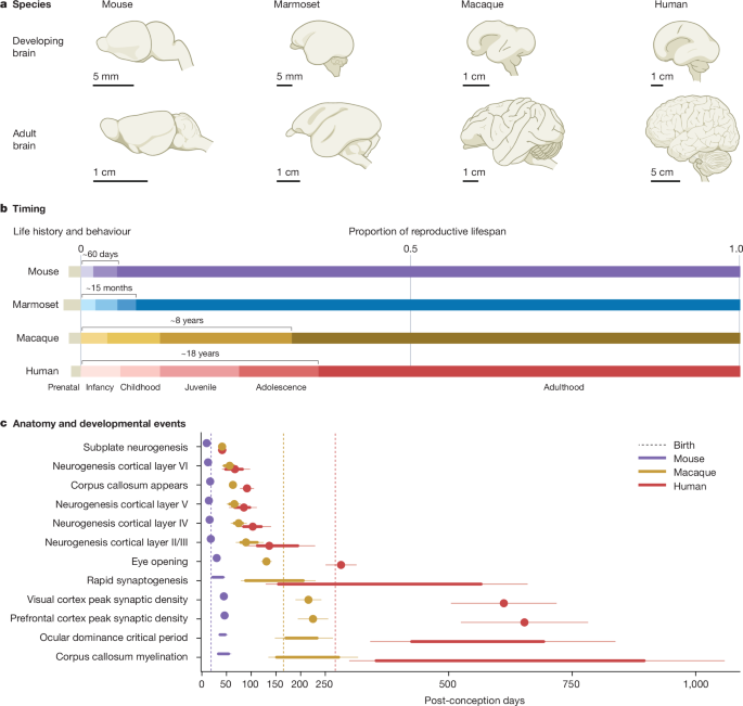
Brust, V., Schindler, P. M. & Lewejohann, L. Lifetime development of behavioural phenotype in the house mouse (Mus musculus). Front. Zool. 12, S17 (2015).
de Castro Leão, A., Duarte Dória Neto, A. & de Sousa, M. B. C. New developmental stages for common marmosets (Callithrix jacchus) using mass and age variables obtained by K-means algorithm and self-organizing maps (SOM). Comput. Biol. Med. 39, 853–859 (2009).
Walker, M. L. & Herndon, J. G. Menopause in nonhuman primates? Biol. Reprod. 79, 398–406 (2008).
Clancy, B., Darlington, R. B. & Finlay, B. L. Translating developmental time across mammalian species. Neuroscience 105, 7–17 (2001).
Workman, A. D., Charvet, C. J., Clancy, B., Darlington, R. B. & Finlay, B. L. Modeling transformations of neurodevelopmental sequences across mammalian species. J. Neurosci. 33, 7368–7383 (2013).

