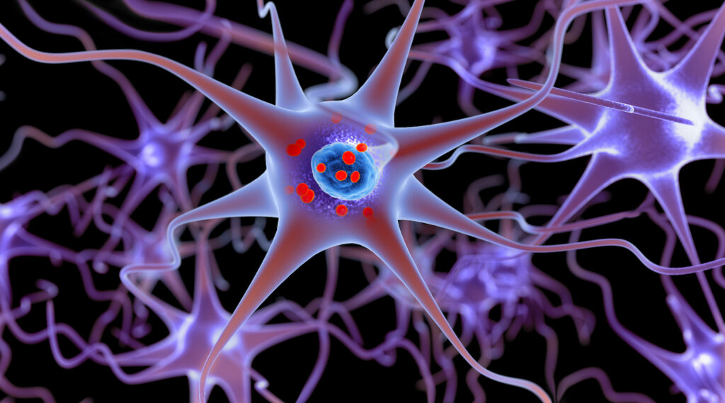
A new study from the Gladstone Institutes has found that a mitochondrial protein, CHCHD2, can drive a sequence of cellular events that lead to the development Parkinson’s disease (PD), findings that show mitochondrial failure may not only be associated with the disease but may initiate it. The research, published in Science Advances, used a mouse model that exhibits the symptoms of a rare, late-onset, inherited form of PD that resembles the most common sporadic form.
“This mouse model provides some of the most compelling evidence to date for how mitochondrial dysfunction can cause typical late-onset Parkinson’s disease,” said senior author Ken Nakamura, MD, PhD, a senior investigator at Gladstone and a professor of neurology at University of California, San Francisco.
The researchers zeroed in on the CHCD2 based on earlier research showing how mutations in it resulted in α-synuclein deposition similar to the more common sporadic form of PD. “The discovery that mutations in the mitochondrial intermembrane space protein CHCHD2… cause an autosomal dominant form of late-onset PD that closely resembles sporadic PD… proves that mitochondrial dysfunction can cause autosomal dominant PD, and strongly implicates it as a causal factor in at least a subset of idiopathic PD,” the researchers wrote.
The challenge of Parkinson’s disease is that has different forms, and even the most common sporadic form comprises an array of disease subtypes driven by both genetic and environmental influences. As is commonly performed for other disease studies, researchers have turned to engineering mouse models of PD to get a better grasp on its different forms and subtypes. Unfortunately, mouse models that carry some of the key mitochondrial mutations linked to PD in humans have failed to develop the tell-tale features of sporadic disease.
Looking for answers to why CHCHD2 mutations might trigger PD, the team developed their own mouse model that knocked in the T61I mutation that is known to be the disease-causing mutation in CHCHD2. They then conducted a comprehensive analysis of the CHCHD2 T61I phenotype in their mouse models and extended observations to brain tissue of patients with sporadic PD.
This method revealed pronounced mitochondrial disruption in substantia nigra dopaminergic neurons, including distorted ultrastructure and CHCHD2 aggregation, as well as disrupted mitochondrial protein-protein interactions in brain lysates, the investigators noted. They also found that mutant CHCHD2 accumulated within mitochondria, which became swollen and altered in structure. The mutated protein also interfered with interactions among respiratory chain proteins, and whole-body metabolic measurements producing a shift from aerobic respiration toward glycolysis. These changes in metabolism occurred before increases in mitochondrial reactive oxygen species (ROS).
“A notable finding was that alpha-synuclein doesn’t accumulate until after levels of reactive oxygen species rise,” said co-first author Szu-Chi Liao, PhD, a former researcher in the Nakamura lab. This activity was in line with the team’s hypothesis that oxidative stress from mitochondrial dysfunction drives α-synuclein aggregation.
To test whether similar mechanisms operate in humans, the researchers collaborated with Australian researchers at the University of Sydney to examine brain tissue from people who had sporadic PD. Their data showed that CHCHD2 protein accumulated in early α-synuclein aggregates in dopamine-producing neurons, a finding that indicates mutant CHCHD2 triggers a sequence of mitochondrial damage, ROS elevation, and α-synuclein pathology that is characteristic of the majority of PD cases.
“This work is a blueprint for how a mitochondrial protein can be disrupted and actually cause Parkinson’s disease,” Nakamura noted.
While these new findings are important in creating a link between CHCHD2 and mitochondrial dysfunction, the researchers noted some limitations of their work including uncertainties about whether CHCHD2 aggregation is a driver of degeneration or part of a compensatory or protective response.
“We propose that the accumulation of CHCHD2 aggregates leads to the disruption of mitochondrial ultrastructure and mitochondrial PPIs. However, an alternate possibility is that a primary disruption of mitochondrial PPIs leads directly to the disruption of energy metabolism, and the aggregation are both secondary processes,” the researchers noted.
Nevertheless, this research could inform future efforts to develop therapeutics for PD. Some approaches suggested by the researchers include targeting ROS, supporting mitochondrial respiration, or modulating metabolic shifts as ways to interrupt disease progression. Along these lines, the Gladstone team now aims to test whether drugs that block ROS and boost cellular energy could stop the chain of events that lead to PD development.

