Researchers headed by a team at the University of Texas at Dallas’ Center for Advanced Pain Studies (CAPS) have made a fundamental discovery about a key mechanism that enables nervous system connections to strengthen.
The discovery, based on studies in mice and on human tissue, centers on phosphorylation, a biochemical process in which a kinase enzyme modifies another protein’s function by adding a phosphate molecule to it. This process is considered fundamental for functions within cells, such as metabolism, structural processes and subcellular signaling.
The findings have direct implications for better understanding the underlying biochemical mechanisms involved in pain, but also potentially in learning and memory, said CAPS director Ted Price, PhD, Ashbel Smith professor of neuroscience in the School of Behavioral and Brain Sciences. The discoveries could also point to potential therapeutic approaches targeting pain.
“This study gets to the core of how synaptic plasticity works—how connections between neurons evolve,” he said. “In this study we found that kinases within the synaptic cleft itself play an important role in synaptic plasticity. These results alter our textbook-level understanding of how synapses work.” The study, Price added, “… has very broad implications for neuroscience.”
Price is a co-corresponding author of the team’s published paper in Science, titled “The synaptic ectokinase VLK triggers the EphB2–NMDAR interaction to drive injury-induced pain.”
The role of extracellular phosphorylation, which occurs outside of cells, isn’t well understood, especially in synapses. “Phosphorylation of hundreds of protein extracellular domains is mediated by two kinase families, but the functional role of these kinases is underexplored,” the team noted.
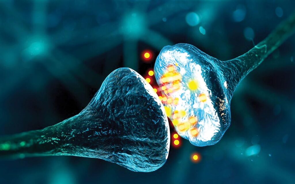
It is into the synapse that a presynaptic neuron releases neurotransmitters, peptides, and proteins that then bind to, activate and regulate receptors on postsynaptic neurons. This information transfer forms the basis of neuronal plasticity, a process that can enhance or diminish the strength of connections between nerve cells involved in learning, memory and pain.
For their newly reported work, Price and colleagues focused on the role that phosphorylating kinases secreted by neurons might play in regulating synaptic signaling. “Extracellular phosphorylation occurs via ectokinases—kinases secreted outside of cells,” explained Price. “It has been known to occur for almost 150 years, but almost nothing has been learned about it in the nervous system.”
The authors further noted, “One example of the importance of extracellular phosphorylation is the interaction between Eph receptor tyrosine kinase B2 (EphB2) and N-methyl-d-aspartate receptor (NMDAR) that occurs at excitatory synapses throughout the brain and spinal cord,” the authors commented. “The EphB-NMDAR interaction is induced by the phosphorylation of a highly conserved extracellular tyrosine residue Y504 in the fibronectin type III (FNIII) domain of EphB2.”
The ectokinase vertebrate lonesome kinase (VLK) was recently shown to have a role in platelet function and bone development. Researchers also knew that phosphorylation via ectokinases was linked to pain but did now know which specific kinase was responsible. They noted that phosphorylation of EphB2 at Y504 promotes NMDAR-dependent pain in mice and is involved in acute postsurgical pain, suggesting an important role for this extracellular phosphorylation within the spinal cord after injury.
For their newly reported research, the scientists specifically “… tested whether a secreted kinase could control the interaction between the receptor tyrosine kinase EphB2 and the N-methyl-d-aspartate receptor (NMDAR), a key regulator of glutamate signaling, and pain.” The studies were headed by teams led by Price and Matthew Dalva, PhD, director of the Tulane Brain Institute and professor of cell and molecular biology at Tulane University.
The results suggest that VLK is also needed for a key interaction between neurons that mediates injury-induced pain. The team discovered that, as the result of an injury, presynaptic neurons secrete VLK, which then phosphorylates the extracellular side of EphB2 extruding from the membranes of postsynaptic nerve cells. This unique process attracts NMDA receptor proteins, which then cluster in the membrane with the EphB2 receptors. NMDA receptors play a key role in learning and memory formation by regulating the electrical potential of neurons to strengthen synaptic connections.
“Increased concentrations of NMDA receptors at the synapse allows for high levels of neuronal activation, leading to greater postsynaptic potentials—a fundamental mechanism of synaptic plasticity,” said Hajira Elahi, PhD, co-first author of the study who completed much of the work as part of her dissertation.
The researchers found that mice genetically engineered to lack VLK in sensory neurons involved in pain did not develop acute hypersensitivity to pain after surgery. “Mice lacking VLK in sensory neurons failed to develop mechanical hypersensitivity following surgical injury but retained normal motor coordination and responses to heat and chemical stimuli,” they pointed out. Conversely, administering VLK to normal mice induced robust pain hypersensitivity that was mediated by NMDA receptor activation. “Intrathecal injection of rVLK induced EphB2–NMDAR interaction and pain-like behaviors, and this effect required NMDAR activity.”
Elahi further reported, “Complementing the mouse studies, we found that human sensory neurons also express and secrete VLK, and that VLK induces the EphB2 and NMDA receptor interaction in human tissue too. This really highlights the translational impact of our work—the ectokinase role of VLK is likely important in human synaptic signaling as well.”
Price said, “We started this project 10 years ago based on our long-standing collaboration with the Dalva lab, initially synthesizing kinases here at UT Dallas. It took us years to get around to testing VLK. When we did, it became very clear that VLK is the kinase that phosphorylates EphB1 and EphB2 receptors, and that VLK activity is sufficient to cause NMDA receptor clustering.”
Co-corresponding author Dalva added, “The finding that neurons release a protein kinase to modify synaptic function is broadly important and suggests many new and unexpected targets. Our work is a great example of collaborative science. The project and our findings would not have been possible without the shared expertise of the different teams participating.”
Although NMDA receptors have long been a potential pain-relief drug target, direct approaches to modulate them are fraught with side effects. “NMDA receptors are involved in almost every aspect of how the nervous system works,” Price said. “Our findings suggest a new way to manipulate NMDA receptors through VLK targeting potentially without huge side effects. In cortical neurons from the brain, VLK seems to be released in an activity-dependent fashion. Because of that, we can envision a model with many implications within the nervous system.”
A potential therapeutic approach to pain relief might include local injections to block VLK in the spine, he said, although more research is needed to determine how widespread the synaptic VLK mechanism is in the nervous system. “We’re most excited about having discovered that kinases act within the synapse, not just inside neurons. It’s a huge update to our understanding of the basic mechanisms that regulate receptors involved in synaptic plasticity,” Price added. “And I think we’re just scratching the surface. Showing that kinase activity within the synaptic cleft is important for how synapses work will have a big impact on how we think about synaptic plasticity.”
In their paper the authors concluded, “Given the essential role of NMDARs in synaptic plasticity and the embryonic lethality of neuronal VLK knockout, this pathway likely extends beyond pain and may offer new therapeutic opportunities for modulating receptor function.”

