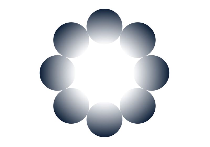
Researchers unraveled part of the neurological circuitry that contributes to illusions.
Illusions are everywhere. For example, the moon appears larger when it rests on the horizon than when it is hanging in the sky. Other visual tricks occur when a person perceives an object in an image that is not actually there, called an illusory contour. Because of the arrangement of the shapes, people may see a square or a triangle on top of a group of circles when neither shape is present.
Scientists hypothesize that this phenomenon arose to identify patterns in a field of view even when part of an object was obscured or the visual information was missing. The mechanisms that reinforce this pattern completion process, though, are unknown.
Researchers at the University of California, Berkeley and the Allen Institute identified neurons that recognize these illusory contours and outlined the circuits that they activate.1 These findings, published in Nature Neuroscience, provide insights about how the brain processes visual information.
Typically, higher visual areas, which integrate and process visual information to enhance object recognition, motion, and other visual tasks, in the visual cortex are responsible for making inferences from information; meanwhile, areas like the primary visual cortex predominantly respond to the specific inputs. However, recent work showed that these high visual areas in mice activate regions in the primary visual cortex, suggesting the possible presence of a circuit between the two areas that promotes these visual inferences.2
Researchers used multi-Neuropixels recordings to image neurons activated by visual input or with optogenetic probe stimulation. This helped them determine where illusory information was encoded in the mouse visual cortex.
Allen Institute
To explore the cells responsible for recognizing the illusory shapes, the team first recorded neuron activity in the mouse primary visual cortex. They identified a subset of neurons that activated in response to the illusory contour. The team then trained a machine learning algorithm to categorize neural activity specific to the recognition of illusory shapes and, using a new set of objects with phantom edges, demonstrated neural activity specific to recognizing these shapes.
Next, to study the potential role of these specialized cells, the team recorded neuron activation in the mouse visual cortex while the animals viewed an image containing a phantom edge. The researchers identified the neurons specific to these illusory shapes and then used optogenetics to activate these same cells without a visual stimulus and captured the neural activation patterns. When they put the activity data into their decoder— even though it was trained on activity from visually-stimulated neurons—it accurately classified the optogenetically-activated neuron network pattern as one that “saw” an illusory contour.
Finally, the researchers demonstrated that these illusion-specific neurons are responsible for completing patterns based on prior experiences and expectations. These neurons reinforce these activity patterns in other neurons, eventually signaling back to the higher visual areas, through feedback loops.
Besides answering questions about how the visual cortex perceives incomplete information, these findings can also inform researchers about conditions like schizophrenia, which is characterized by hallucinations. “If you don’t understand how those objects are formed and a collective set of cells work together to make those representations emerge, you’re not going to be able to treat it; so understanding which cells and in which layer this activity occurs is helpful,” said Jérôme Lecoq, a neuroscientist at the Allen institute and study coauthor, in a press release.

