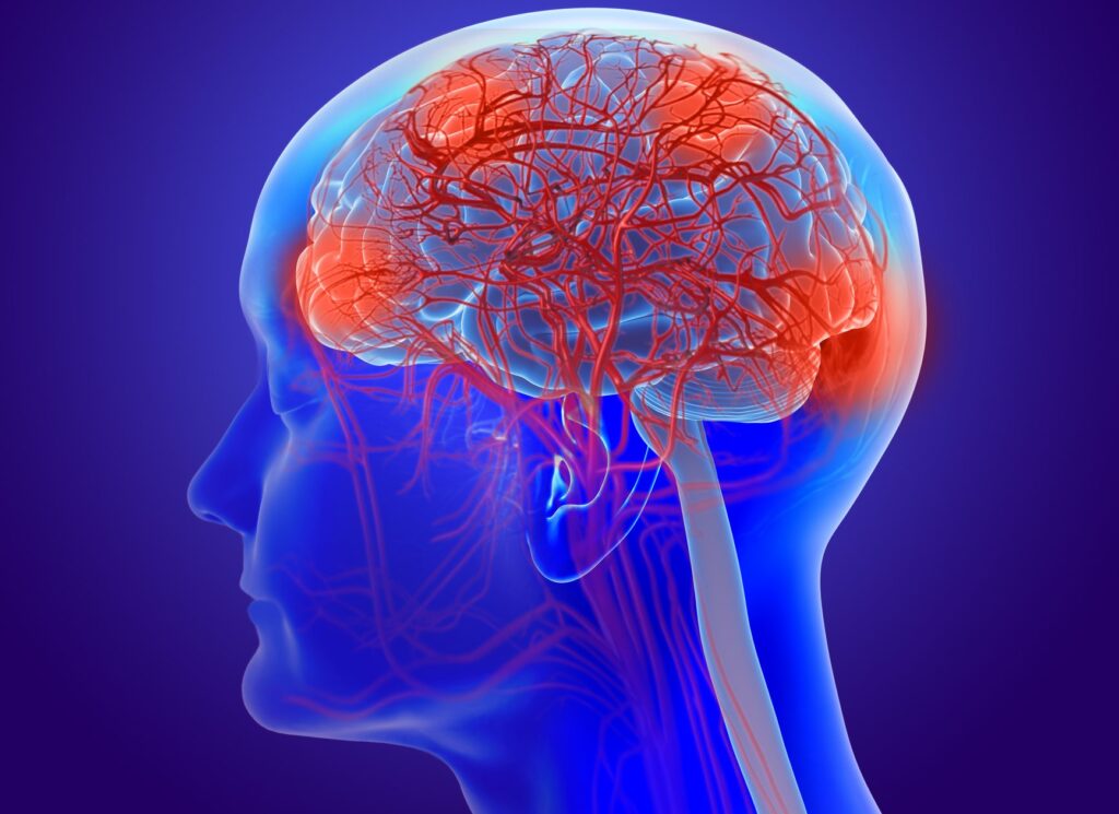
Researchers at the University of Southern California (USC) have developed a noninvasive method to measure the rhythmic pulsations of the brain’s smallest blood vessels, which was impossible until now in live human brains. In a study published in Nature Cardiovascular Research, the new technique revealed a link between an increase in pulsations in the brain’s microvessels and signs of aging and neurodegeneration, especially in deep white matter regions.
“Arterial pulsation is like the brain’s natural pump, helping to move fluids and clear waste,” said Danny J. J. Wang, PhD, professor of neurology and radiology at the Keck School of Medicine of USC and senior author of the study. “Our new method allows us to see, for the first time in people, how the volumes of those tiny blood vessels change with aging and vascular risk factors. This opens new avenues for studying brain health, dementia, and small vessel disease.”
While it has long been known that the stiffness and pulsations of large arteries is linked to a higher risk of stroke, dementia, and small vessel disease, there were no methods to measure these pulsations in the smallest blood vessels found in the human brain. All methods available until now were invasive and therefore used only in animal studies.
To overcome this limitation, Wang and colleagues combined two existing magnetic resonance imaging (MRI) techniques, allowing them to track subtle volume changes in the brain’s microvessels with high resolution. The new method combines vascular space occupancy (VASO) MRI, a non-invasive technique to detect changes in the brain’s blood volume, with arterial spin labeling (ASL) MRI, a technique used to quantify the movement of blood throughout the brain.
“Being able to measure these tiny vascular pulses in vivo is a critical step forward,” said Arthur W. Toga, PhD, director of the Stevens Neuroimaging and Informatics Institute at the Keck School of Medicine. “This technology not only advances our understanding of brain aging but also holds promise for early diagnosis and monitoring of neurodegenerative disorders.”
The new technique was used to measure microvascular pulsations in the brains of 11 younger adults and 12 older adults. Results showed a significant increase in pulsatility in the brains of older participants, especially within specific white matter areas known to be high-risk zones for a variety of neurological disorders.
“These findings provide a missing link between what we see in large vessel imaging and the microvascular damage we observe in aging and Alzheimer’s disease,” said Fanhua Guo, PhD, a postdoctoral researcher in Wang’s research group and lead author of the study.
Previous research points at a link between neurodegeneration and the brain’s glymphatic system, which is responsible for clearing waste such as the build up of beta amyloid proteins that characterizes Alzheimer’s disease. Increased vascular pulsatility had formerly been linked to an impairment of the glymphatic system, but until now these measurements could only be made indirectly or in animal models.
Going forward, the group is interested in exploring how to adapt this method for clinical use, possibly using lower resolution MRI scanners and reducing the time it currently takes to perform the procedure. Future work will also look at whether changes in vascular pulsatility could be used as a predictive biomarker for early interventions in Alzheimer’s and other related conditions.
“This is just the beginning,” said Wang. “Our goal is to bring this from research labs into clinical practice, where it could guide diagnosis, prevention, and treatment strategies for millions at risk of dementia.”

