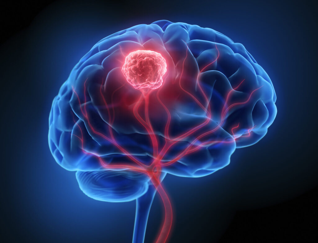
An AI tool developed by a Harvard Medical School–led team of researchers accurately distinguishes between glioblastoma and other central nervous system (CNS) tumors during surgery, helping surgeons make critical treatment decisions in real time.
The tool, known as PICTURE (Pathology Image Characterization Tool with Uncertainty-aware Rapid Evaluations), demonstrated almost perfect accuracy in differentiating glioblastoma from primary central nervous system lymphoma (PCNSL), a far less common malignancy that is frequently misdiagnosed as glioblastoma.
“I was struck by the very high level of accuracy,” said study senior author Kun-Hsing Yu, MD, PhD, associate professor of biomedical informatics in the Blavatnik Institute at HMS and HMS assistant professor of pathology at Brigham and Women’s Hospital.
“Distinguishing CNS tumors is an exceptionally complex task, and even seasoned experts can find it challenging to reach the correct diagnosis,” Yu told Inside Precision Medicine. “PICTURE’s strong performance in this context underscores both the strength of its design and the potential of AI to support clinical decision-making.”
There are more than 100 distinct CNS tumor subtypes, the most common of which is glioblastoma. Patients with glioblastoma are typically treated with surgical removal of the tumor, yet their median survival is limited to around eight months. PCNSL has overlapping pathology features with glioblastoma, but the treatment is different, and the median survival is longer, at three years.
If PCNSL is found during surgery initially performed to remove a tumor, further surgical intervention is typically discontinued to preserve neurological function, and patients are instead referred for radiotherapy combined with chemotherapy.
“Thus, accurate distinction between glioblastoma and PCNSL at both intraoperative and final diagnostic stages is essential to avoid unnecessary surgery and ensure timely initiation of appropriate therapy,” write Yu and co-authors in Nature Communications.
To address the diagnostic challenge, Yu and team developed the PICTURE system using 2141 formalin-fixed paraffin-embedded (FFPE) and frozen section CNS tissue pathology slides collected from five medical centers and worldwide.
“What makes PICTURE unique is that it was designed to capture the wide spectrum of CNS tumor pathology, drawing not only from individual datasets but also from patterns reported in the medical literature,” Yu explained. “In addition, it is uncertainty-aware: unlike many AI models that produce an answer even when the supporting evidence is weak, PICTURE can recognize when a case lies outside what it has learned and flag that uncertainty. This combination of broad representation and the ability to ‘know when it doesn’t know’ makes it more reliable and safer for clinical use than conventional AI approaches.”
When the researchers tested the performance of PICTURE on FFPE samples from patients not used in the AI training process, they found that it could distinguish glioblastoma from PCNSL with 99.8% accuracy. The impressive performance was replicated in five independent cohorts with both FFPE and frozen samples and was significantly better than some state-of-the-art computational pathology methods currently available for use in the field.
PICTURE also outperformed nine human neuropathologists with a range of clinical experience. In representative tests that included 20 frozen section slides and 20 FFPE slides, each with diverse morphological features of glioblastoma and PCNSL, the evaluators misclassified PCNSL as glioblastoma in 38% of cases. Of note, among the 113 misdiagnosed instances across evaluators, 72% were associated with evaluator-reported low to moderate diagnostic confidence (≤50%) and several participants noted in post-evaluation feedback that the task was “more challenging than expected,” highlighting the complexity of CNS tumor evaluation.
Further work showed that Yu and team’s uncertainty quantification approach meant that PICTURE was able to identify 67 types of rare central nervous system cancers that were neither gliomas nor lymphomas and were not seen in the training phase of the model.
The authors explain that standard machine learning methods are often trained to classify outcomes into pre-defined categories, which results in patients being pigeonholed into one of the prespecified diagnostic groups.
“By flagging cases likely to be neither glioblastoma nor PCNSL, [PICTURE] enables clinicians to promptly identify rare or atypical presentations that warrant further evaluation, thereby enhancing the trustworthiness of computer-aided diagnostics,” they write. “These results suggested that PICTURE could facilitate routine pathology diagnosis, which requires clinicians to accurately identify common cancers while staying vigilant for rare diseases and atypical pathology manifestations.”
Despite the positive findings, Yu said there are still two critical hurdles to overcome before PICTURE can be adopted widely in routine practice: regulatory approval and integration.
“As with all medical AI systems, formal clearance will be required before it can serve as a primary tool for clinical decision-making,” he remarked. Then, PICTURE “must be embedded directly into pathology workflows so results are delivered seamlessly and on time, without disrupting routine practice.”
Yu concluded that “the key impact of PICTURE is its ability to shift diagnosis and treatment decisions from manual interpretation to a more systematic, data-driven process.”
He added: “For precision medicine, this translates into faster and more accurate diagnosis for tumor types that may look similar under the microscope but require very different treatment strategies, teal-time intraoperative diagnostic support to guide surgical decision-making at the point of care, and identification of ambiguous cases, ensuring they are directed to confirmatory testing rather than risking misclassification.
“In short, PICTURE could strengthen both the precision and personalization of care for CNS cancer patients by directly linking tumor morphology to underlying biology and clinical outcomes.”

