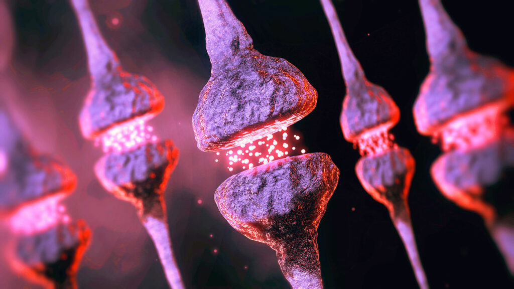
For decades, neuroscientists have debated whether synaptic vesicles “kiss-and-run” or do an irreversible “full collapse” when releasing neurotransmitters. Now, a team in China has frozen that moment in time—literally—and revealed a third act in the dance of neuronal communication.
Cracking the code of neurotransmission
Our brains rely on fast, precisely timed synaptic transmissions between neurons. At the heart of this process is synaptic vesicle (SV) exocytosis—the release of neurotransmitters triggered by an electrical signal, or action potential. But despite its central role, the structural and biophysical mechanics of SV release have remained elusive. Was it a fleeting “kiss-and-run” or a full-on “collapse” into the presynaptic membrane? The answer has evaded scientists for over half a century, and a team of researchers at the University of Science and Technology of China (USTC) set out to understand this mechanism through a newly developed method.
Published in Science, the study titled “Kiss-shrink-run unifies mechanisms for synaptic vesicle exocytosis and hyperfast recycling,” introduces a new biophysical model that reconciles the long-standing dichotomy. SV release, it turns out, is neither purely transient nor wholly irreversible—it’s a hybrid process with a critical shrinking phase.
Understanding this mechanism isn’t just academic—it’s foundational. The fidelity and efficiency of synaptic transmission underpins neuronal communication—everything from learning and memory to neurodegenerative disease. A clearer picture could unlock new therapeutic avenues and deepen our grasp of brain function. The team of researchers at the University of Science and Technology of China (USTC) set out to understand this mechanism and developed a time-resolved, cellular cryo-electron tomography (cryo-ET) platform. By combining optogenetic stimulation, or using light to trigger neural activity, with high-speed plunge-freezing, they captured over 1,000 tomograms of intact excitatory synapses at intervals from 0 to 300 milliseconds post-action potential.
This millisecond-scale imaging allowed the team to reconstruct the full timeline of SV exocytosis and rapid recycling. Within just 4 milliseconds, vesicles formed a ~4 nm fusion pore (“kiss”), then shrank to half their original surface area (“shrink”). By 70 milliseconds, most were recycled via the “run” pathway, while others underwent “full-collapse” fusion with the presynaptic membrane.
A new mechanism: “kiss-shrink-run”
This “kiss-shrink-run” mechanism not only explains the structural basis for rapid and reliable neurotransmission but also opens new doors for studying synaptic plasticity and brain function and disorders. The cryo-ET technique itself sets a new standard for in situ analysis of membrane dynamics and molecular interactions with unmatched spatiotemporal resolution.

